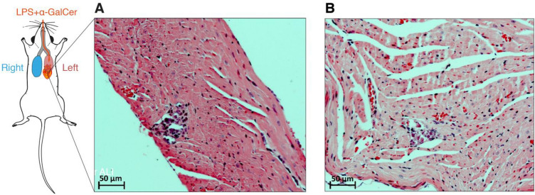
Fig. 3.
Fragments of myocardium of the right ventricle of male ICR mice in the DAD model on days 7 (A) and 14 (B) of the follow-up: small foci of necrosis of cardiomyocytes at stages of mononuclear infiltration (on the left) and organization (on the right) as indirect signs of volume overload of the right heart, and pulmonary hypertension. Staining with hematoxylin and eosin. Magnification: 200x

