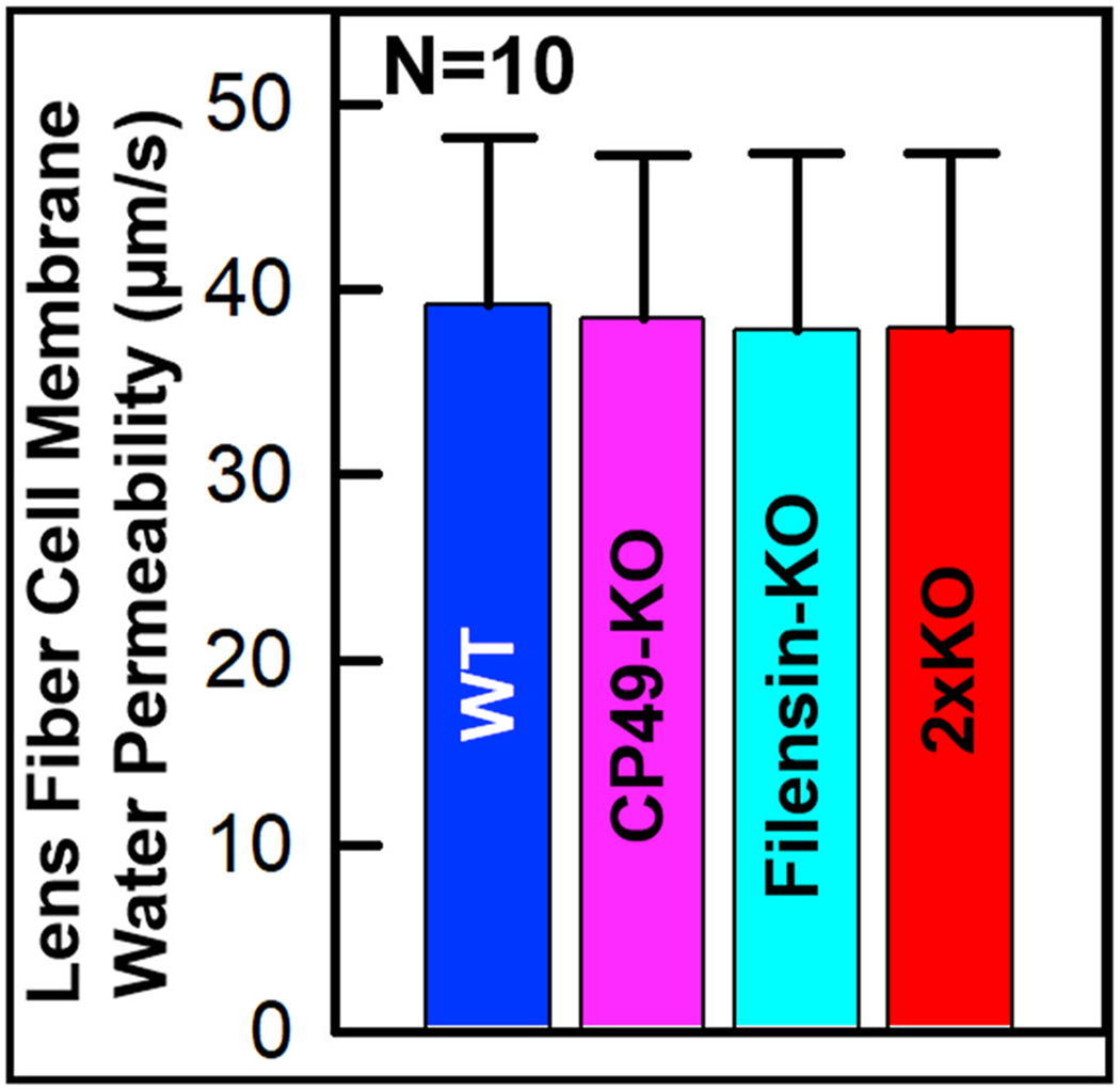
Fig. 7.

Lens fiber cell membrane water permeability. Membrane vesicles from WT, CP49-KO, filensin-KO and 2xKO lens fiber cells were tested. Each bar represents mean ± SD. SE values: WT: 2.8394; CP49-KO: 2.8016; filensin-KO: 3.0067; 2xKO: 2.9719. Fiber cell membrane vesicles exhibited no statistically significant alteration (P > 0.05) in Pf compared to those of WT. Ten vesicles and four mice were used per study.
