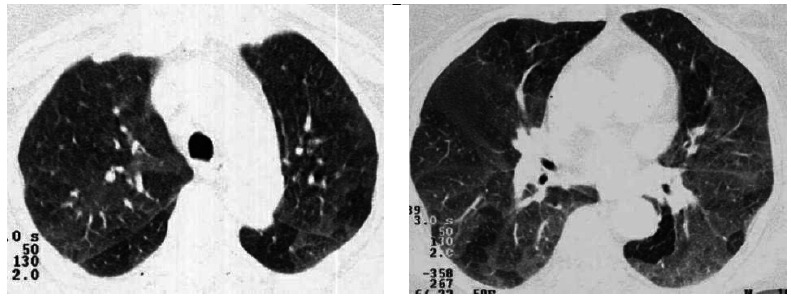
Figure 1. Axial CT scans with lung window settings acquired in the expiratory phase, revealing, in the posterior segments of the lower lobes, different parenchymal densities, with areas of decreased attenuation associated with air trapping due to small airway obstruction.

