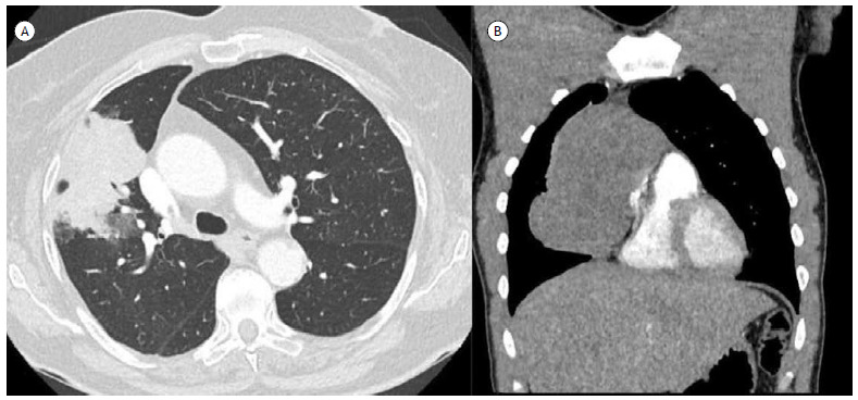
Figure 11. Axial CT scans with lung window settings. In A, a mass with lobulated contours in the anterior segment of the upper lobe of the right lung in a patient with lung adenocarcinoma. In B, an anterior mediastinal (prevascular) mass, with soft-tissue attenuation, in a patient with mediastinal seminoma.

