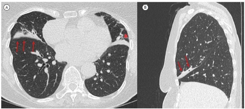
Figure 2. In A, an axial CT scan with lung window settings revealing atelectasis of the right middle lobe (arrows) and lingula (asterisk). In B, a coronal CT scan with lung window settings showing a slight displacement of the oblique fissure, bronchi, and adjacent vessels (arrows).

