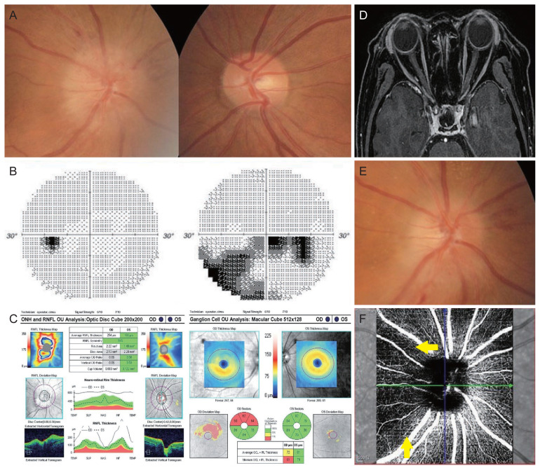
Fig. 1.
Nonarteritic ischemic optic neuropathy in the right eye after vaccination against Coronavirus disease 2019. (A) Disc photographs at presentation showing a swollen disc with several splinter peripapillary hemorrhages in the right eye and a normal disc with cupping (not a disc-at-risk) in the fellow eye. (B) Inferior arcuate and cecocentral visual field defects respecting the horizontal meridian in the right eye. (C) Optical coherence tomography showing thickened peripapillary retinal nerve fiber layer (RNFL) and thinning of macular retinal nerve fiber layer, corresponding to the visual field defect. (D) Gadolinium-enhancing T1-weighted magnetic resonance imaging of the brain and orbits showing normal optic nerves in both eyes. (E) Disc photograph after high-dose steroid treatment showing a progression of atrophy in the supero-temporal sector of the right optic disc. (F) Optical coherence tomography angiography 4 months after vaccination, indicating a decreased peripapillary capillary density adjacent to the superior and temporal sectors of optic disc (indicated by yellow arrows). The patient provided written informed consent for publication of the research details and clinical images. ONH = optic nerve head; OU = both eyes; OD = right eye; OS = left eye; C/D = cup/disc; TEMP = temporal; SUP = superior; NAS = nasal; INF = inferior; GCL = ganglion cell layer; IPL = internal plexiform layer.

