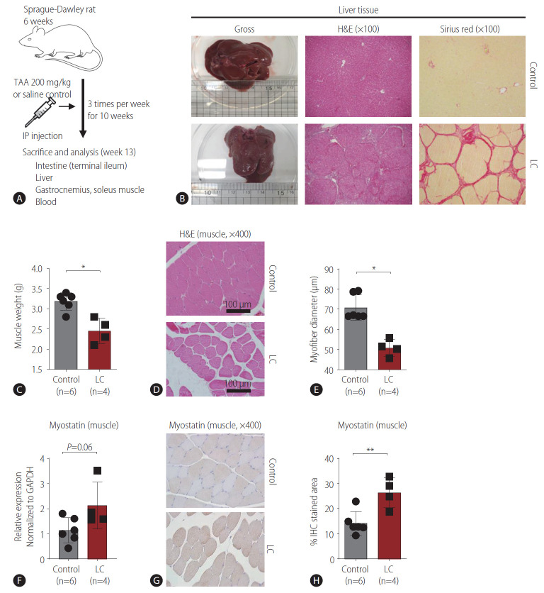
Figure 1.
Rat model of liver cirrhosis (LC) and sarcopenia. (A) Summary of the rat model of LC. Six-week-old Sprague-Dawley rats were intraperitoneally administered thioacetamide (200 mg/kg) or saline control three times per week for 10 weeks, and tissues from each organ of interest and blood were collected at 13 weeks. (B) Examples of gross, hematoxylin and eosin (H&E)-stained, and Sirius red-stained livers from the control and LC groups. (C) Comparison of calf muscle weights in the control (n=6) and LC (n=4) groups. (D, E) Comparison of myofiber diameters in the control (n=6) and LC (n=4) groups. H&E staining (D) and the graph (E). (F) Comparison of myostatin expression in the calf muscles of control (n=6) and LC (n=4) rats were measured by RT-PCR. (G, H) Comparison of myostatin staining in the calf muscles of control (n=6) and LC (n=4) rats were measured by immunohistochemistry (IHC). Histological findings (G) and the graph (H). TAA, thioacetamide; IP, intraperitoneal. *P<0.05. **P<0.01.

