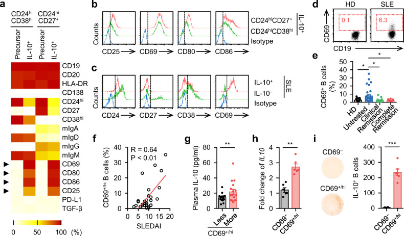
Fig. 2. Phenotypic characteristics of IL-10-secreting Breg cells in SLE.
a, b FACS analysis of the phenotypic characteristics of the untreated CD24hiCD38hi and CD24hiCD27+ B cells (precursor), as well as IL-10+ cells derived from CD24hiCD38hi and CD24hiCD27+ B cells by CPG-DNA induction (a). Representative histogram of CD25, CD69, CD80, and CD86 expression were shown in b (n = 5). c Expression of CD24, CD27, CD38, and CD69 in IL-10+ B cells purified from untreated SLE patients was detected by FACS (n = 6). d, e FACS analysis of CD69+/hi B cells from healthy donors and untreated SLE patients. Representative FACS plots were shown in d. The portions of CD69+/hi B cells relative to total B cells from 21 healthy donors, 30 untreated SLE patents, 14 clinical remission SLE patients, and 12 complete remission SLE patients were shown in e. f, g Associations of circulating CD69+/hi B cells with disease progression (f) and plasma level of IL-10 from untreated SLE patients (g) (n = 30). h, i Expression of IL-10 by CD69− and CD69+/hi B cells isolated from untreated SLE patients were detected by PCR (h) and ELISpot (i), respectively (each n = 6). Data were acquired from more than four independent experiments and shown as means ± SEM. Pearson’s correlation analysis for f. Significance was determined with one-way ANOVA test (e), Student’s t test (g–i). *p < 0.05, **p < 0.01, ***p < 0.001

