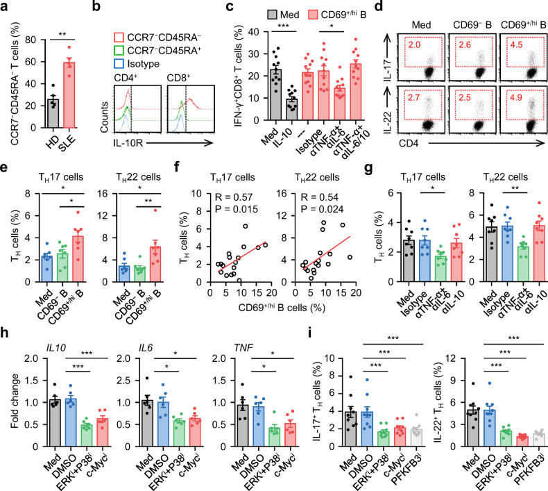
Fig. 6. Targeting c-Myc-elicited glycolysis abrogates inflammatory Breg cell mediated pathogenic response.
a, b FACS analysis of CCR7−CD45RA− effector memory cells T cells in circulating T cells (a) and IL-10R expression (b) on effector memory T cells in blood samples from 5 healthy donors and 5 untreated SLE patients. c Purified T cells from SLE patients were cultured in medium, treated with recombinant human IL-10, or cocultured with CD69+/hi B cells in the presence of isotype control, anti-TNF-α antibody plus anti-IL-6 antibody, or anti-TNF-α antibody plus anti-IL-6 antibody plus anti-IL-10 antibody for 7 d. IFN-γ expression in CD8+ T cells was examined using FACS (n = 11). d, e Purified T cells from SLE patients were left untreated or cultured with CD69− B cells or CD69+/hi B cells for 7 d. Differentiation of TH17 and TH22 was determined by FACS (n = 7). Representative dotplots and statistical data were shown in d and e, respectively. f Associations of circulating CD69+/hi B cells with circulating TH17 and TH22 cells in untreated SLE patients (n = 17). g Purified T cells from untreated SLE patients were cultured with autologous CD69+/hi B cells in the presence of isotype control, anti-TNF-α antibody plus anti-IL-6 antibody, or anti-IL-10 antibody for 7 d. Differentiation of TH17 and TH22 was determined by FACS (n = 8). h, i Purified CD69+/hi B cells from SLE patients were cultured in medium or pretreated with DMSO, inhibitor against c-Myc, PFKFB3, or ERK plus P38. Thereafter, the cells were incubated with autologous T cells for 7 d. Cytokine expression in B cells (h) and differentiation of TH17 and TH22 (i) was measured by real-time PCR and FACS (n = 6 for h; n = 9 for i). Data were acquired from more than four independent experiments and shown as means ± SEM. Pearson’s correlation analysis for f, significance was determined with Student’s t test (a) or one-way ANOVA test (c, e, g–i). *p < 0.05, **p < 0.01, ***p < 0.001

