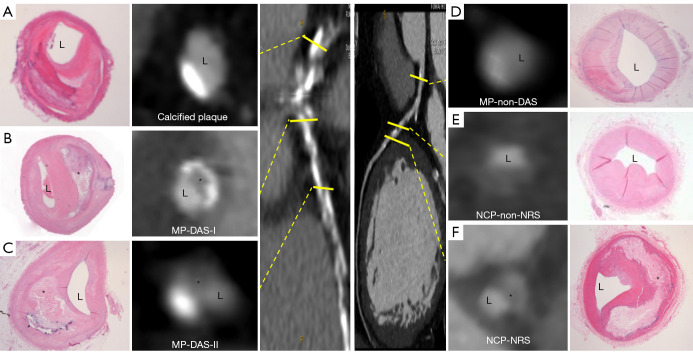
Figure 2.
Plaque classifications as assessed by CCTA in vivo. CCTA of an in vivo donor heart with the anatomical position of the plaque cross sections. The plaque-calcified-pattern included calcified (A), MP-DAS-type I (B), MP-DAS-type II (C), MP-non-DAS (D), NCP-NRS (E), NCP-non-NRS (F). The corresponding histology slides: (A) pure calcified; (B) calcified with fibrous cap thickness <65 µm and lipid core accounting for >40% of the plaque’s total area-type I; (C) calcified with fibrous cap thickness <65 µm and lipid core accounting for >40% of the plaque’s total area-type II; (D) calcified and fibrous plaque; (E) pathological intimal thickening; (F) late fibroatheroma with fibrous cap thickness <65 µm and lipid core accounting for >40% of the plaque’s total area. *, necrotic core. L, lumen; MP, mixed plaque; DAS, diamond-attenuation-sign; NCP, non-calcified plaque; NRS, napkin-ring-sign; CCTA, coronary computed tomography angiography.

