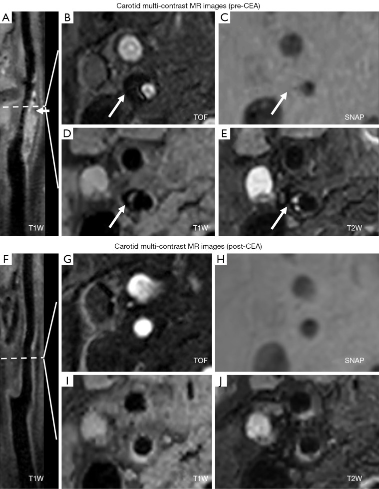
Figure 2.
A 76-year-old male patient had a calcific plaque in the right internal carotid artery and underwent revascularization pre- and post-CEA. The patient’s scores of pre-MoCA and post-MoCA were 17 and 20, respectively. Calcification can be seen in the multi-contrast MR images (white arrows). (A) 3D T1W curved planar reconstruction image before CEA; (B) 3D TOF image before CEA; (C) SNAP image before CEA; (D) 2D T1W image before CEA; (E) 2D T1W image before CEA; (F) 3D T1W curved planar reconstruction image after CEA; (G) 3D TOF image after CEA; (H) SNAP image after CEA; (I) 2D T1W image after CEA; and (J) 2D T1W image after CEA. CEA, carotid endarterectomy; MR, magnetic resonance; MoCA, Montreal Cognitive Assessment; T1W, T1-weighted; TOF, time-of-flight; SNAP, simultaneous non-contrast angiography intraplaque hemorrhage; T2W, T2-weighted.

