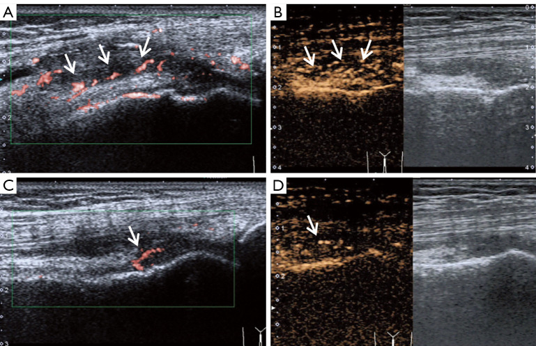
Figure 4.
Longitudinal dorsal ultrasound of the knee joint. (A,B) In the active period of RA, superb microvascular imaging (SMI) shows a small amount of color signal in the hypertrophic synovium (Grade 2), and contrast-enhanced ultrasound (CEUS) exhibits intensive enhancement (Grade 3). (C,D) In RA in the clinical remission period, strip blood flows are still visible on both SMI and CEUS (Grade 1). Arrow point to the vessels.

