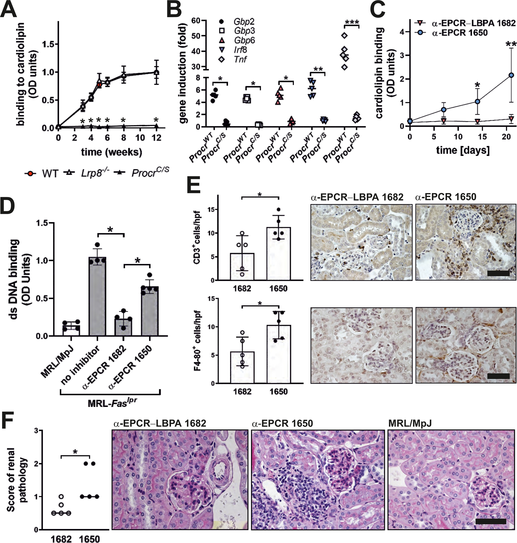
Figure 6: EPCR–LBPA signaling drives aPL expansion and autoimmune pathology in vivo.

(A) Mice of the indicated genotypes were immunized with aPL HL5B and anti-cardiolipin titer determined at the indicated times; n=10, *P<0.0001 for ProcrC/S vs. WT; one-way ANOVA. (B) IgG isolated 12 weeks after the start of aPL immunization were used to stimulate human MM1 cells for 1 hour for induction of the indicated genes; n=5, *P<0.05, **P=0.011, ***P<0.0001; two-way ANOVA, Sidak’s multiple comparisons test. (C) MRL-Faslpr lupus-prone mice were treated with the indicated α-EPCR antibodies at an age of 4 weeks (day 0) and anti-cardiolipin titers were determined in serum at the indicated time points; n=5, *P=0.03; **P<0.0001; two-way ANOVA, Sidak’s multiple comparisons test. (D) Antibodies to double stranded (ds) DNA were measured in α-EPCR 1650- and α-EPCR–LBPA 1682-treated MRL-Faslpr mice 2 weeks after the last dose or in 6-week-old MRL/MpJ control or MRL-Faslpr mice; n=4–5, *P<0.0001; one-way ANOVA. (E) Immune cell infiltration of α-EPCR-treated MRL-Faslpr mice; n=5, *P<0.025. (F) Renal pathology scores of α-EPCR-treated MRL-Faslpr mice; n=5, *P=0.0317; Mann–Whitney U test. Scale bars=80 µm.
