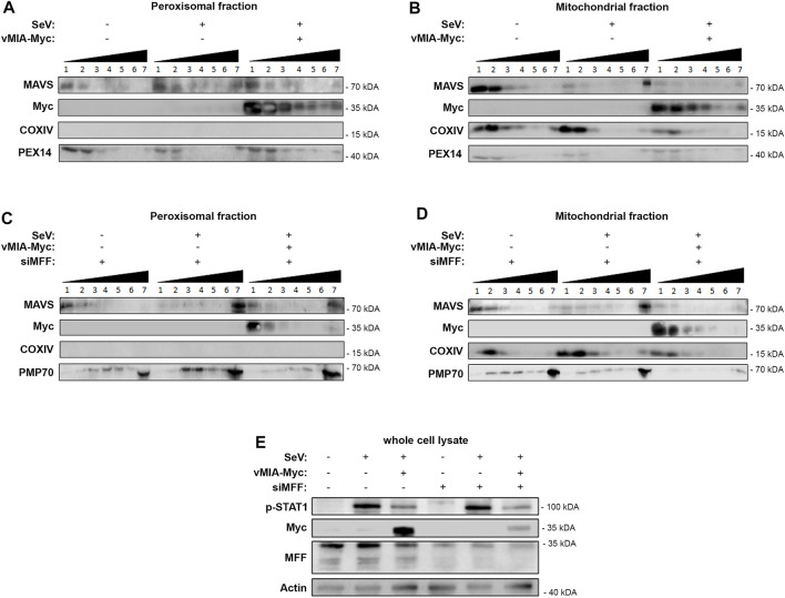
FIGURE 5.
vMIA inhibits MAVS oligomerization at peroxisomes and mitochondria. MFF is essential for the vMIA-mediated inhibition of MAVS oligomerization at peroxisomes but not at mitochondria. (A,B) HEK293T cells infected with SeV in the presence or absence of vMIA. Density gradient assay was performed to demonstrate the separation of endogenous MAVS based on its density. 1—7 represent the fractions isolated from the gradient assay, where 1 represents the fraction with lowest density and 7 represents the fraction with highest density. (A) Peroxisome-enriched fraction, (B) Mitochondria-enriched fraction. (C,D) HEK293T cells infected with SeV in the presence or absence of vMIA and in the absence of MFF. Density gradient assay was performed to demonstrate the separation of endogenous MAVS based on its density. 1—7 represent the fractions isolated from the gradient assay, where 1 represents the fraction with lowest density and 7 represents the fraction with highest density. (C) Peroxisome-enriched fraction, (D) Mitochondria-enriched fraction. (A–D) Immunoblots were performed with antibodies against MAVS, Myc-tag, COXIV, PEX14 and PMP70. (E) Whole cell lysates were resolved by SDS-PAGE. SeV infection, and consequential activation of MAVS downstream signalling, was confirmed using anti-p-STAT1. vMIA-Myc overexpression and MFF silencing were also confirmed using anti-Myc and anti-MFF, respectively. Antibody against Actin was used as loading control.

