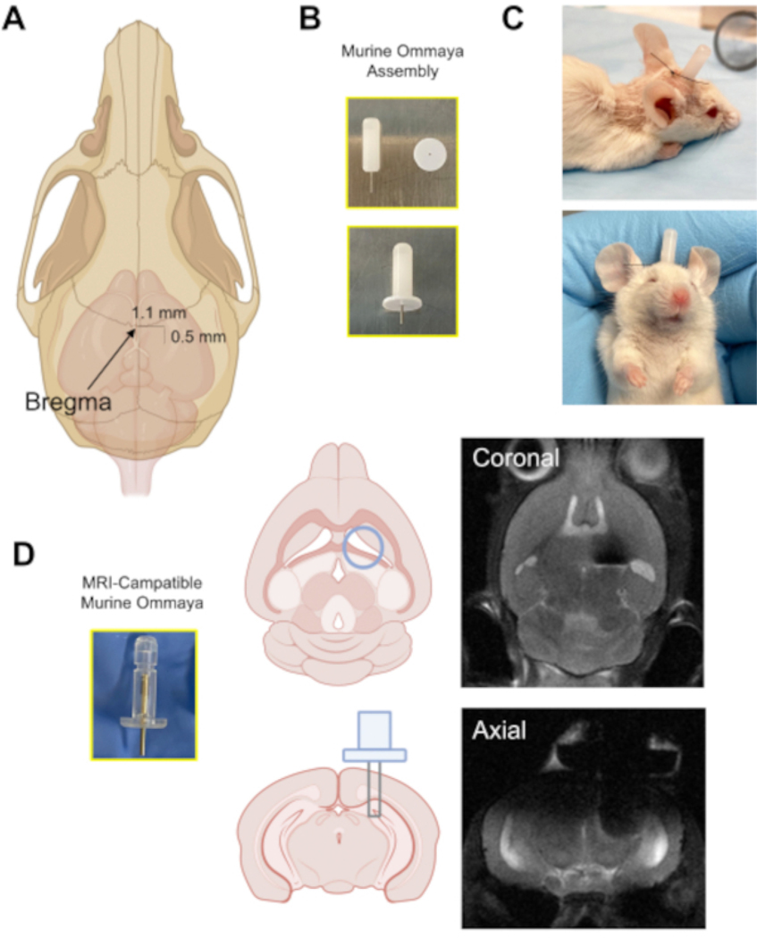
Figure 3: The implantation of the Murine Ommaya device.
(A) An illustration with an arrow pointing at the location of the bregma on the skull, and the approximate distance at which a burr hole is drilled in the skull (0.5 mm posterior/1.1 mm lateral) from the bregma using a microdrill. (B) A Murine Ommaya is assembled by combining a metal cannula and a 1 mm spacer as the base for glue attachment to the skull. (C) Representative images of mice that had Murine Ommayas implanted; these mice are monitored to ensure they are bright, alert, and reactive before receiving any injections. (D) An example of the prototype magnetic resonance imaging-compatible Murine Ommaya and representative brain magnetic resonance imaging images of Murine Ommaya implants.

