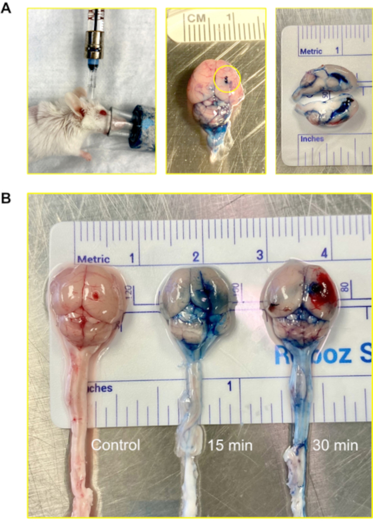
Figure 4: Intraventricular (central nervous system) injection using the Murine Ommaya.

(A) An image of an injection, accessing the ventricle and central nervous system via the Murine Ommaya. Mice remain under anesthesia during the injection. In the example, the Murine Ommaya is connected to the miniature port that is attached to a prefilled Hamilton syringe. Injections are performed using an automatic injection set at an infusion rate of 1 μL/min and a volume of 5–7 μL. An image of a mouse brain injected with Evans Blue is shown. The circle shows where the Murine Ommaya was attached. No leakage of the dye was observed on the exterior of the brain. A cross section of the brain shows the lateral ventricles were filled with the dye; the dye did not penetrate brain parenchyma. (B) Images of mouse brains after 15 and 30 min following the injection of Evans Blue dye. The dye infiltrated the brain (15 min) and began to circulate on the spinal cord (30 min). Out of 5 mice, 4 received dye for visualization, and 1 served as control. The experiment was repeated in triplicate.
