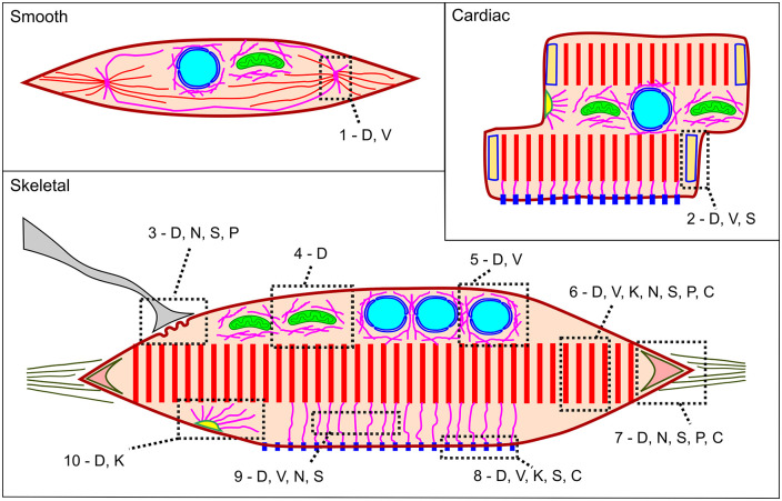
Figure 6.
Scheme showing the distribution of intermediate filaments in smooth, cardiac and skeletal muscle: D: desmin, V: vimentin, K: cytokeratin, N: nestin, S: synemin, P: paranemin, C: syncoilin, 1: dense body, 2: intercalated disk, 3: neuromuscular junction, 4: perimitochondrial, 5: perinuclear, 6: sarcomere, 7: myotendinous junction, 8: costamere, 9: cytoplasm, 10: desmosome. We do not depict other regions where IF proteins have been described, such as cytoplasm and sarcolemma because these categories are ambiguous. (A color version of this figure is available in the online journal.)

