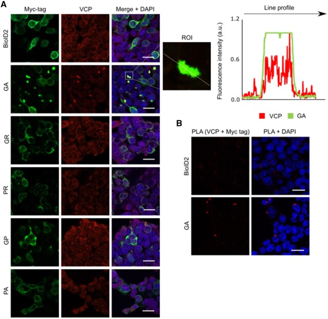
Figure 3.
Cellular localization of DPRs and VCP in HEK293 cells. (A) HEK293 cells transfected with the DPR constructs, revealed using an anti-Myc antibody (green) and endogenous VCP (red). VCP co-localized with polyGA, but not with the other DPRs. The line profile plot indicates the intensity distribution of the green and red channels through the white line in the magnified view of the region of interest (ROI) in the merged panel (middle). Scale bars = 20 µm. (B) Representative proximity ligation assay (PLA) using anti-Myc and anti-VCP antibodies revealed interactions between polyGA and VCP (red dots).

