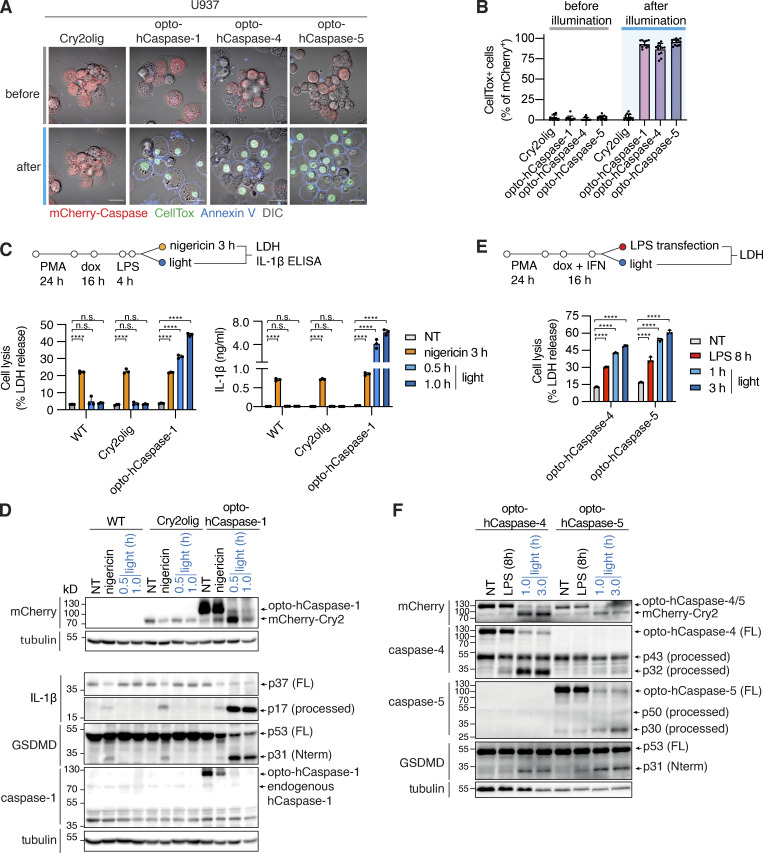
Figure 2.
Optogenetic pyroptosis induction in human macrophage-like cell lines. (A and B) Representative images and quantification of CellTox+ PMA-differentiated U937 cells expressing the indicated constructs before and after 1 h of blue light illumination. Scale bar, 20 µm. (C and E) LDH and IL-1β release from WT U937 or U937 cells expressing the indicated constructs, treated with nigericin (C), transfected with LPS (E), or illuminated with blue light. NT, non-treated. (D and F) Immunoblots of combined cell lysates and supernatants from C or E, respectively. Anti-mCherry is used for full-length and processed opto-hCaspase-1/-4/-5 and Cry2olig. A and C–F are representative of at least three independent experiments, each with three technical replicates per experiment (at least n = 9), B is pooled from three experiments, each with quadruplicate technical replicates (n = 12). Mean ± SD, ****, P < 0.0001 (one-way ANOVA). Source data are available for this figure: SourceData F2.

