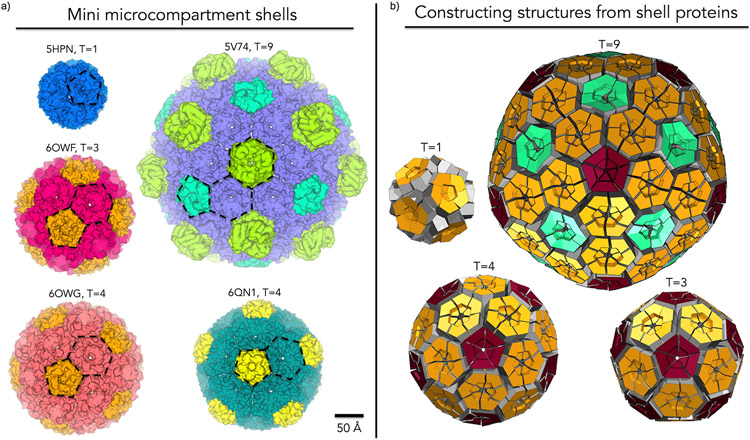
Figure 4:
Gallery of miniaturized MCP shells highlighting diversity in shape, size and number of components. (a) First columns (top to bottom): PDB: 5HPN – Shell from an engineered circularly permuted BMC shell protein, PduA, which formed a pentamer; PDB: 6OWF – Shell constructed from one BMV and one BMC-H from a beta-carboxysome, T=3; PDB: 6OWG – Shell constructed from one BMV and one BMC-H from a beta-carboxysome, T=4. Second column (top to bottom): PDB: 5V74 – 6.5 MDa shell constructed from one BMV, one BMC-H and three types of presently indistinguishable BMC-T proteins from a Haliangium ochraceum MCP. PDB: 6QN1 – mini GRM2 shell constructed from one BMV and three presently indistinguishable BMC-Hs. (b) Geometric models representing different icosahedral triangulation patterns observed. BMV (red), BMC-H (orange), and BMC-T(aquamarine). In the T=1 case an engineered BMC-H protein rearranged to form pentameric units.

