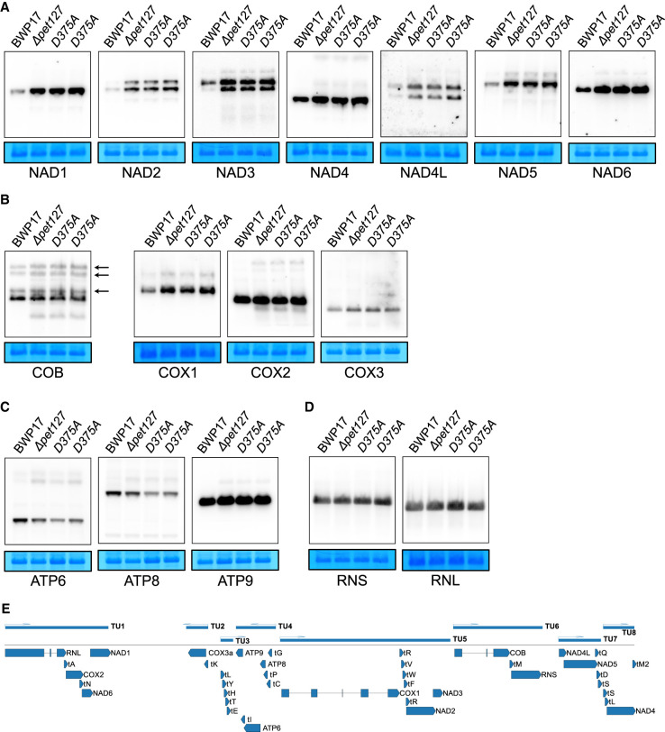
FIGURE 3.
Changes in steady-state RNA levels in ΔCapet127 and Capet127D375A mutant strains. Northern blot analysis of mitochondrial mRNA and rRNA transcripts from wild-type (BWP17), homozygous ΔCapet127 (ΔCapet127), mutant, and Capet127D375A (D375A) catalytic mutants (two independent strains). (A) mRNAs encoding subunits of Complex I. (B) mRNAs encoding subunits of Complex III (COB) and Complex IV (COX). Arrows indicate the splicing intermediates of COB. (C) mRNAs encoding subunits of the ATP synthase (Complex V). (D) rRNAs of the small (RNS) and large (RNL) subunits of the mitoribosome. RNAs were prepared from purified mitochondria and separated by agarose/formaldehyde gel electrophoresis in denaturing conditions. Methylene blue staining of the small subunit mitochondrial rRNA in the blot is shown below each autoradiogram as a loading control. In panels A–D, blot series [NAD3, RNS]; [NAD4, COB]; [RNL, COX1]; [COX2, NAD5] were prepared by stripping and rehybridizing the same membrane, hence the same loading controls. (E) Schematic map of C. albicans mtDNA (without the second identical repeat region) showing the location of genes in primary transcription units (TU), according to Kolondra et al. (2015).

