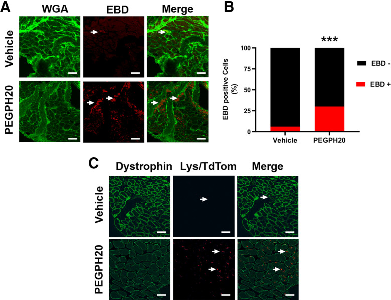
Figure 3.
PEGPH20 disrupts of skeletal muscle architecture that is accompanied by macrophage infiltration. A, Muscle extracellular membrane (green, WGA) is disrupted 3 d after the administration of PEGPH20. EBD (red) usually remains in intravascular spaces in intact tissue but after hyaluronidase administration it leaks into the affected skeletal muscle. B, Percentage of myofibers positive for EBD 3 d after PEGPH20 administration. C, At the same time point macrophages (red, LysM/tdTomato) infiltrate the connective tissue surrounding the skeletal muscle in contrast to the almost nonpresent macrophages in the vehicle-treated animals. White scale bar: 50 μm. ***p < 0.001 χ2 test.

