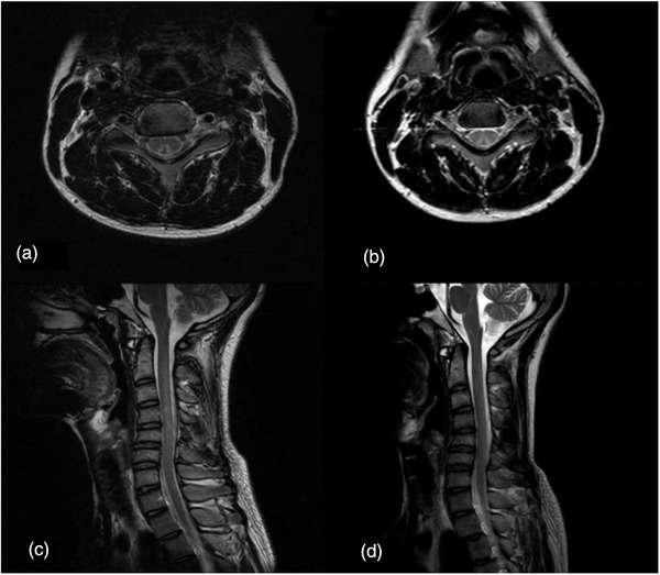
FIGURE 3.

(a, c) Magnetic resonance image (MRI) examination of patients with nitrous oxide abuse at initial consultation showed increased T2 signal in the spinal cord at the C3–C6 level. (b, d) MRI reexamination after 1 month showed reduction of increased T2 signal in the spinal cord at the C3–C6 level
