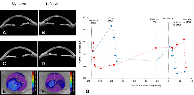
Figure 1.
Composite showing: (A) swept-source optical coherence tomography (SS-OCT) of the right eye post-Descemet membrane endothelial keratoplasty (DMEK); (B) SS-OCT of the left eye post-DMEK; (C) SS-OCT of the right eye following DMEK graft rejection; (D) SS-OCT of the left eye following DMEK graft rejection; (E) pachymetry map of the right eye following DMEK graft rejection; (F) pachymetry map of the left eye following DMEK graft rejection; (G) graph demonstrating corneal thickness of the right eye (red) and the left eye (blue) over time after vaccination. The timeline of procedures is shown, including: DMEK, re-DMEK and posterior capsulotomy with neodymium-doped yttrium aluminum garnet laser (YAG).

