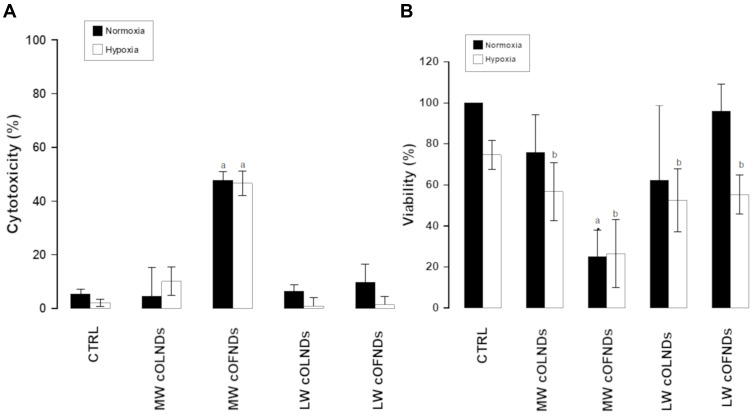
Figure 2.
cND cytotoxicity and effects on human keratinocyte viability, under normoxic and hypoxic conditions. HaCaT cells (3x 10⁵ cells/mL for LDH studies and 1.6×10⁵ cells/mL for MTT studies) were left untreated or treated with MW or LW cOLNDs and cOFNDs for 24 h in normoxia (20% O2, black columns) or hypoxia (1% O2, white columns). After collection of cell supernatants and lysates, cytotoxicity percentage was measured through LDH assay (A) and cell viability percentage was measured through MTT assay (B). Results are shown as means ± SEM from three independent experiments. Data were also evaluated for significance by ANOVA. Vs normoxic control cells: a p<0.001; vs hypoxic control cells: b p<0.05.

