PLOS One logoLink to PLOS One
. 2022 Apr 18;17(4):e0266688. doi: 10.1371/journal.pone.0266688

Serum Amyloid A is not obligatory for high-fat, high-sucrose, cholesterol-fed diet-induced obesity and its metabolic and inflammatory complications

Ailing Ji 1,2, Andrea C Trumbauer 2, Victoria P Noffsinger 1,2, Hayce Jeon 3,4, Avery C Patrick 3, Frederick C De Beer 1,2,5, Nancy R Webb 3,2,5, Lisa R Tannock 1,2,5,6, Preetha Shridas 1,2,5,*
Editor: Michael Bader7
PMCID: PMC9015120  PMID: 35436297

Abstract

Several studies in the past have reported positive correlations between circulating Serum amyloid A (SAA) levels and obesity. However, based on limited number of studies involving appropriate mouse models, the role of SAA in the development of obesity and obesity-related metabolic consequences has not been established. Accordingly, herein, we have examined the role of SAA in the development of obesity and its associated metabolic complications in vivo using mice deficient for all three inducible forms of SAA: SAA1.1, SAA2.1 and SAA3 (TKO). Male and female mice were rendered obese by feeding a high fat, high sucrose diet with added cholesterol (HFHSC) and control mice were fed rodent chow diet. Here, we show that the deletion of SAA does not affect diet-induced obesity, hepatic lipid metabolism or adipose tissue inflammation. However, there was a modest effect on glucose metabolism. The results of this study confirm previous findings that SAA levels are elevated in adipose tissues as well as in the circulation in diet-induced obese mice. However, the three acute phase SAAs do not play a causative role in the development of obesity or obesity-associated adipose tissue inflammation and dyslipidemia.

Introduction

Obesity is associated with a wide variety of pathologies, collectively known as metabolic diseases resulting in significant morbidity and mortality. Chronic and low-grade inflammation is a hallmark of obesity and key factor for the development of obesity comorbidities [1]. Obesity-driven adipose tissue inflammation has been extensively characterized and its role in the development of insulin resistance and impaired glucose metabolism leading to the development of type 2 diabetes have been established [2, 3]. However, the obesity-induced factors responsible for the inflammatory state in adipose tissue and the relationship of dysregulated adipose tissue to systemic inflammation still remain unclear.

Serum amyloid A (SAA) is a family of acute phase proteins whose circulating levels rise (up to 1,000-fold or more) in an acute phase response. There are three inducible SAA subtypes that likely arose through gene duplication [4]. Humans express two acute-phase SAA proteins, SAA1 and SAA2 which are 96% homologous over their entire length, and correspond to mouse SAA1.1 and SAA2.1. In addition, mice encode a third conserved acute-phase SAA gene, Saa3. Saa3 is considered to be a pseudogene in humans due to an early stop codon [57]. The liver is believed to be the predominant source of circulating SAA during an acute inflammatory state and in chronic inflammatory diseases such as rheumatoid arthritis and lupus, although we [8] and others [9, 10] have shown that all three SAA isoforms are also significantly induced in adipose tissue in endotoxemic mice. Yet another isoform of SAA, SAA4, is expressed in humans and mice and is synthesized constitutively [11]. Unlike the three acute-phase isoforms, SAA4 does not show massive induction during acute inflammation [7, 11]. Chronic and modest elevations in SAA concentrations have been demonstrated in obesity, metabolic syndrome or diabetes [1214], and circulating concentrations of SAA correlate with body fat [15]. Weight loss tends to decrease these levels [1518]. In a prospective population-based study, a statistically significant association between systemic SAA levels and the development of type 2 diabetes was observed in an elderly western European population that was independent of various other established type 2 diabetes risk factors [19]. However, though SAA levels are positively correlated with both obesity and diabetes, whether there is any physiological relevance to this association is not clear. Several in vitro and selected in vivo studies demonstrated SAA to invoke inflammatory properties and functions that would be expected to promote the development of obesity, inflammation and insulin resistance. Studies with recombinant SAA have indicated that SAA acts as a chemoattractant for both monocytes and polymorphonuclear cells [20]. When not associated with high density lipoprotein (HDL), SAA is known to increase the production of cytokines, reactive oxygen species and nitric oxide [2124]. The results from in vitro studies using recombinant SAA are questionable now as differences between recombinant SAA and endogenous SAA purified from acute-phase plasma have been found [25]. Our studies using SAA purified from mouse plasma shows activation of NLRP3 inflammasome activation in mouse macrophage cells [26]. A previous report indicated that SAA induced hypoxia, a common event associated with fat expansion [27].

Earlier studies in mice indicated that deficiency of SAA3 blunts weight gain induced by obesogenic diet, hyperlipidemia and adipose tissue specific inflammation and macrophage accumulation are attenuated in female but not in male mice [28]. Ahlin et al. studied the role of human SAA1 overexpression in adipose tissues of mice, and observed no significant impact on diet-induced obesity, adipose tissue inflammation and insulin resistance [29]. A study by de Oliveira et al. [30] showed that suppression of SAA in male Swiss Webster mice by SAA-1 and 2 -targeted antisense oligonucleotides caused a significant reduction in adipose tissue expansion, expression of inflammatory markers, macrophage infiltration into the adipose tissues and remarkable improvement in glucose and insulin tolerance in mice fed a high fat diet. It is not yet clear whether the SAA isoforms compensate for the deficiency of each other and whether deficiency of all SAA subtypes impacts obesity-associated inflammation and metabolic dysfunction. Although it is now widely accepted that increased adiposity leads to elevated SAA in adipose tissue and serum in both humans and mice, evidence that SAA plays a key role in obesity-associated metabolic dysfunction is somewhat lacking. Whether SAA plays a functional role in the development of obesity and/or obesity-induced adipose tissue inflammation, insulin resistance and other metabolic complications, or is merely a consequence of adipose tissue inflammation and thus a marker of obesity remains an unresolved question. SAA is involved in the development of several chronic inflammatory diseases, including atherosclerosis [31, 32] and angiotensin II-induced abdominal aortic aneurysm formation [33]. Here, we investigated whether SAA plays a causal role in the development of diet-induced obesity and associated metabolic changes in mice.

Materials and methods

Animals

C57BL/6 mice deficient in SAA1.1, SAA2.1 and SAA3 (TKO) mice were generously provided by Drs. June-Yong Lee and Dan Littman, New York University. The TKO mice were generated by inserting a premature stop codon into exon 2 of saa3 in the SAA1.1/2.1-deficient (SAAKO) mouse [33, 34] using CRISPR-Cas9 technology as described previously [35, 36]. The details of the genomic organization of the SAA1.1 and SAA2.1 genes and the construction of SAAKO mice were described earlier [32]. Genotyping to identify SAA TKO mice is performed using a multi-step approach. The first step confirmed targeting of the SAA1.1 and SAA2.1 loci utilizing a 3-primer PCR reaction as described earlier [37]). The second step to validate SAA3 targeting is performed by two sets of PCRs as described in S1A Fig. All mice were provided food and water ad libitum. Animals were housed in micro-isolator cages and maintained on a 14-hr light/10-hr dark cycle. All studies were performed in accordance with the Public Health Service Policy on Humane Care and Use of Laboratory Animals with the approval of the University of Kentucky Institutional Animal Care and Use Committees.

Diet and study design

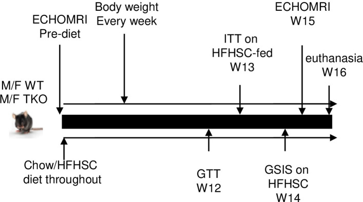
The study design is depicted in Fig 1. Briefly, 17-weeks old male and female C57BL/6 (WT) and TKO mice were fed either normal chow (18% calories from fat, 58% from carbohydrate and 24% from protein) or a high fat, high-sucrose diet with 0.15% added cholesterol (HFHSC, F4997, Bioserv, Flemington, NJ) ad libitum for 16 weeks. The diet regimen was selected based on previous studies, where it significantly induced SAA expression in adipose tissues and plasma, and increased macrophage accumulation in adipose tissues [28, 38]. The HFHSC diet provides 20.5% of calories as protein, 36% as fat and 36% as carbohydrate. Body weight was measured weekly and body composition was measured by NMR spectroscopy (Echo MRI).

Fig 1. Study design.

Fig 1

Intraperitoneal glucose tolerance test (GTT)

Mice were fasted for 4 h and then given intraperitoneal (i.p.) injection of d-(+)-glucose (Sigma-Aldrich, St. Louis, MO) at 1.5 g/kg body weight. Blood glucose was measured in tail vein blood (Contour Blood glucose monitoring system) just prior to injection of glucose (time 0) and at 30, 60, 90, 120, 150 and 180 min after glucose administration.

Intraperitoneal insulin tolerance test (ITT)

Mice were fasted for 4 h, and then given human insulin (Novolin R) i.p. at 1.0 IU/kg body wt. Blood glucose was measured at 0, 30, 60, 90, 120, and 150 min post injection from tail vein prick. None of the mice developed hypoglycemic shock (glucose levels below 20 mg/dL) and unresponsive to touch as a result of hypoglycemia.

Glucose-stimulated insulin secretion

Glucose-stimulated insulin secretion (GSIS) was performed as described earlier [39]. Briefly, mice were fasted for 16 h, anesthetized, and then blood samples were collected from the retro-orbital sinus before and 15 min after intraperitoneal glucose injection (3 g/kg). Insulin levels in the plasma samples were determined.

Plasma SAA measurements

Plasma SAA concentrations were determined using a mouse SAA ELISA kit (Tridelta Development Ltd, Maynooth, County Kildare, Ireland). Plasma SAA3 concentrations were determined using a mouse SAA3 ELISA kit (Millipore Sigma, Burlington, MA).

Plasma lipid analyses

Plasma total cholesterol, free fatty acid, triglyceride and HDL concentrations were measured using enzymatic kits (Wako Chemicals, Richmond VA.).

Liver triglyceride and cholesterol quantitation

Triglycerides from liver samples were measured according to a previously published method [40]. Briefly, ~50 mg of liver tissue was homogenized in 1 ml of chloroform-methanol (2:1 v/v). The homogenate was then diluted 1:10 with chloroform-methanol (2:1 v/v) and 10 μl of the diluted homogenate was evaporated. The evaporated sample was resuspended directly in 200 μl of Triglycerides Reagent (Pointe Scientific, Canton, MI) and the assay was performed following the manufacture’s description. Cholesterol assay was performed following Folch method [41] with modifications. Briefly, similar to triglyceride assay, ~50 mg of liver tissue was homogenized in 1 ml of chloroform-methanol (2:1 v/v). The homogenate (50 μl) was evaporated and resuspended in 1% Triton X-100 in water. Aliquots were assayed for cholesterol content using a colorimetric kit (Wako Chemicals, Richmond VA).

RNA isolation and quantitative RT-PCR

Total RNA was isolated from mouse adipose tissues according to the manufacturer’s instructions (Qiagen, Valencia, CA). RNA samples were incubated with DNase I (Qiagen, Valencia, CA) for 15 min at RT prior to reverse transcription. Adipose tissue RNA (0.25–0.5 μg) was reverse transcribed into cDNA using the Reverse Transcription System (Applied Biosystems, Waltham, MA). After 4-fold dilution, 5 μl was used as a template for real-time RT-PCR. Amplification was done for 40 cycles using Power SYBR Green PCR master Mix Kit (Applied Biosystems, Waltham, MA). Quantification of mRNA was performed using the ΔΔCT method and normalized to GAPDH. The primers used for the quantification of SAA mRNA were designed to recognize all three inducible isoforms of mouse SAA, SAA1.1, SAA2.1 and SAA3 (Forward: GACATGTGGCGAGCCTAC and reverse: TTGGGGTCTTTGCCACT). In some studies, SAA3 mRNA abundance was specifically quantified using primers specific for SAA3 mRNA (Forward: TTTCTCTTCCTGTTGTTCCAGTC and reverse: TCCCAATGTGCTGAATAAATACTTGTGA). Other primer sequences will be provided on request.

Western blotting

Adipose tissues from the experimental mice were homogenized in RIPA buffer (Sigma-Aldrich, St. Louis, MO) containing a protease inhibitor cocktail (Sigma-Aldrich, St. Louis, MO). The homogenate (50 μg protein) was electrophoresed on a 4–50% polyacrylamide gradient gel (Bio-Rad, Hercules, CA) and immunoblotted with a mixture of two antibodies (rabbit anti-mouse SAA1 + SAA2 antibody, 1: 1000 dilution, Cat number ab199030; abcam, Cambridge, UK) and rabbit anti-mouse SAA3 (a gift from Dr. Phillip Scherer, University of Texas Southwestern) to identify all three acute-phase SAA isoforms, SAA1.1, SAA2.1 and SAA3. The specificity of each of these antibodies has been validated in a previous study [8].

Histology

Gonadal fat tissues were collected from the experimental mice and fixed in 10% formaldehyde, paraffin embedded, cut into 5-μm sections, and stained with hematoxylin (Vector laboratories, Burlingame, CA). Adipocyte area was quantified for 3 randomly selected sections from 3 mice from each group of experimental mice using Nikon NIS-elements software.

Statistics

Data are expressed as mean ±SEM. Results were analyzed by Student’s t test or one-way analysis of variance followed by Sidak’s multiple comparison test. Values of p<0.05 were considered statistically significant.

Results

SAA expression is induced with HFHSC diet feeding

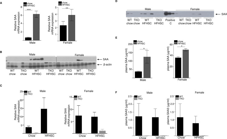
To investigate whether SAA plays a role in the development of diet-induced obesity and insulin resistance, male and female wild type (WT) and SAA-deficient (TKO) mice were fed either normal rodent diet or a high fat, high sucrose diet with added cholesterol (HFHSC) ad libitum for 16 weeks (study design in Fig 1). At study termination, total SAA mRNA abundance in gonadal adipose tissue was significantly increased in both male (9-fold increase) and female (1.6-fold increase) WT mice fed HFHSC diet compared to the chow fed mice of the same sex (Fig 2A). SAA protein was detected by immunoblotting in adipose tissue of WT mice fed HFHSC, but not chow diet (Fig 2B). As expected, SAA protein was not detected in the adipose tissues of SAA-TKO mice fed either chow or HFHSC diets. There was a trend for increased SAA mRNA expression in the livers of WT mice fed HFHSC diet compared to chow, however the difference was not significant in either male or female mice (Fig 2C). As expected, SAA mRNA was undetectable in livers of TKO mice (Fig 2C).

Fig 2. SAA expression is significantly induced in the adipose tissues of obese mice.

Fig 2

A) SAA mRNA abundance in gonadal adipose tissue from chow and HFHSC fed WT male (left panel) and female (right panel) mice after 16 weeks of diet feeding (n = 5 mice/group) B) Adipose tissue lysates (50 μg protein/lane) obtained from WT and TKO mice at the end of the study were immunoblotted for SAA (top panel) and β-actin as loading control (bottom panel). SAA proteins were undetectable in the adipose tissues of chow fed WT mice and all TKO mice. C) SAA mRNA abundance in liver tissue from chow and HFHSC fed WT and TKO male (left panel) and female (right panel) mice after 16 weeks of diet feeding (n = 4–5 mice/group). D) Plasma SAA in WT and TKO mice at the end of the study by western blot. 1 μl plasma was used/lane. E) SAA levels by elisa in chow and HFHSC fed WT male and female mice after 16 weeks of diet feeding. F) plasma SAA3 levels by elisa in chow and HFHSC fed WT male and female mice after 16 weeks of diet feeding. Data are mean ±SEM; * = P ≤ 0.05, ** = P ≤ 0.01 and *** = P ≤ 0.001.

SAA was detected by western blot in the plasmas of WT male mice fed either chow or HFHSC diet, however in WT female mice, SAA was barely detectable only in the HFHSC fed mice (Fig 2D). As expected, SAA was undetectable in both chow or HFHSC diet fed male or female TKO mice (Fig 2D). Consistent with the western blot analysis, analysis of plasma SAA (SAA1.1 and SAA2.1, the predominant forms of acute-phase SAA in circulation, [8]) by elisa indicated significant increase after HFHSC diet feeding compared to chow in both male (3.2-fold increase) and female (1.4-fold increase) WT mice (Fig 2E), consistent with increased systemic SAA in response to obesogenic diet. It was of interest to assess the specific induction of SAA3 after HFHSC feeding, since we [8] and others [42] previously reported that SAA3 is a major SAA isoform expressed in mouse adipose tissue during inflammation. Compared to plasma SAA1.1 and SAA2.1levels (Fig 2E; up to 200 μg/ml), SAA3 levels in the plasma were modest (Fig 2F; ~0.2–4.0 μg/ml), consistent with our earlier observation in mice injected with lipopolysaccharide [8]. There was no diet-induced increase in SAA3 in both male and female WT mice, as expected, SAA3 was undetectable in the plasma of all the TKO mice used in the experiment (Fig 2F). Consistently, SAA3 mRNA abundance was significantly increased in the adipose tissues of male and female WT mice fed HFHSC diet compared to the chow-fed mice of the same sex, whereas SAA3 mRNA was at or below the level of detection in adipose tissues of all TKO mice (S1B Fig). The expression of the constitutively expressed SAA, SAA4 [43] was not significantly different in adipose tissues among the different groups of experimental mice (S1C Fig). SAA3 mRNA expression was also not significantly altered in the livers of WT mice after HFHSC diet feeding compared to chow (S1D Fig).

Deficiency of SAA does not affect the development of diet-induced obesity

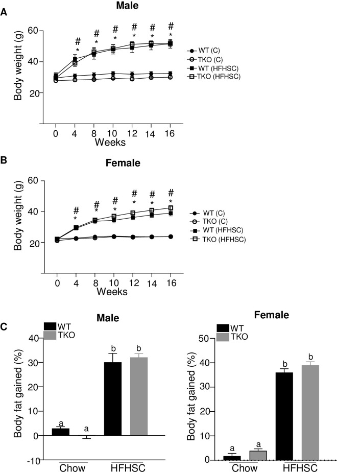
Compared with chow-fed mice, both male and female mice fed the HFHSC diet gained significantly more weight over the course of the study (Fig 3A and 3B), predominantly due to an increase in fat mass as measured by EchoMRI (Fig 3C). There was no significant difference in body weight or fat gain between the genotypes for either males or females after HFHSC diet feeding, indicating that the loss of SAA did not impact the development of obesity (Fig 3A–3C).

Fig 3. Deficiency of SAA does not affect the development of diet-induced obesity.

Fig 3

A) body weight changes in male mice during the course of diet feeding B) body weight changes in female mice during the course of diet feeding C) percentage body fat gained in 15 weeks of chow and HFHSC diet feeding in male (left panel) and female mice (right panel). n = 5–15 mice/group; Data are mean ±SEM; A and B: * and # = P ≤ 0.05 between WT (C) and WT (HFHSC) and between TKO (C) and TKO (HFHSC) respectively. In C, groups that are not significantly different (p ≥ 0.05) are indicated with the same letter.

Deficiency of SAA in HFHSC-fed mice has modest effects on glucose homeostasis

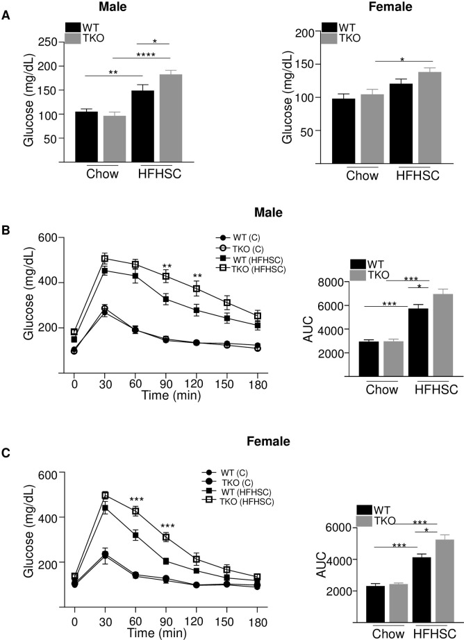
HFHSC diet caused a significant increase in fasting glucose levels in both WT and TKO male mice compared to chow diet (Fig 4A). HFHSC diet-fed male TKO mice had significantly increased fasting glucose levels than WT male mice fed the same diet (Fig 4A, left panel). In female mice, HFHSC diet feeding significantly increased fasting glucose levels only in TKO mice and not in WT mice compared to chow fed mice of the same genotype (Fig 4A, right panel). To investigate whether SAA deficiency impacts diet-induced glucose intolerance in the absence of alterations in adiposity, intraperitoneal glucose tolerance tests (GTT) were performed in male and female WT and TKO mice 12 weeks after initiation of diet feeding. For mice fed the normal rodent diet there was no impact of genotype on glucose excursions in either male or female mice (Fig 4B and 4C). As expected, mice fed HFHSC diet showed significantly impaired glucose tolerance compared to the corresponding chow fed mice, irrespective of their strain or sex (Fig 4B and 4C). Compared to WT mice, there was a modest but significantly increased impairment in glucose tolerance in both male (Fig 4B) and female (Fig 4C) TKO mice after HFHSC diet feeding.

Fig 4. Loss of SAA has a significant but modest effect on diet-induced glucose tolerance.

Fig 4

A) Basal glucose levels in male and female WT and TKO mice after 12 weeks of chow and HFHSC diet feeding (n = 5–15). Data are mean ±SEM; * = P ≤ 0.05; ** = P ≤ 0.01 and *** = p ≤ 0.001. B) intraperitoneal glucose tolerance test (IPGTT) was performed in male mice after 12 weeks of chow and HFHSC diet feeding (n = 10/group). The area under the curve (AUC) is shown in the right panel. Data are mean ±SEM; ** = P ≤ 0.01 between WT HFHSC and TKO HFHSC group of mice. * = P ≤ 0.05 and *** = P ≤ 0.001. C) IPGTT was performed in female mice after 12 weeks of chow and HFHSC diet feeding (n = 5-15/group). The area under the curve (AUC) is shown in the right panel. Data are mean ±SEM; *** = P ≤ 0.001 between WT HFHSC and TKO HFHSC group of mice. * = P ≤ 0.05 and ** = P ≤ 0.01.

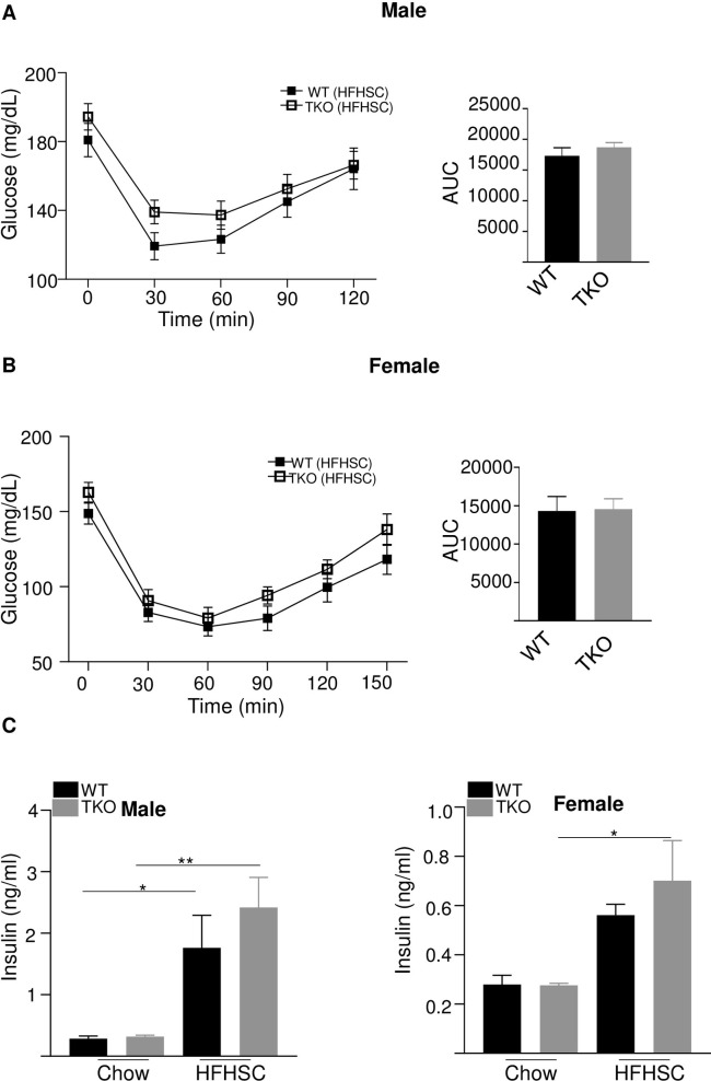
To assess insulin sensitivity, the ability of insulin to decrease blood glucose concentration was determined by performing insulin tolerance tests (ITT) after 13 weeks of HFHSC feeding. There was no significant difference in insulin sensitivity with HFHSC diet in both male and female WT and TKO of the same sex (Fig 5A and 5B). There was also no difference in insulin tolerance between WT and TKO male (S2 Fig left panel) or female (S2 Fig right panel) mice fed chow diet (S2 Fig). Fasting insulin levels were significantly higher in HFHSC diet-fed WT and TKO male mice than that of chow-fed mice of the same strain and gender (Fig 5C, left panel). However, in female mice, HFHSC diet feeding for 14 weeks significantly increased fasting insulin levels only in TKO and not in WT mice compared to the chow-fed mice of the same strain (Fig 5C, right panel).

Fig 5. Loss of SAA does not affect insulin tolerance or plasma fasting insulin levels.

Fig 5

A) insulin tolerance test (ITT) was performed in male WT and TKO mice after 13 weeks HFHSC diet (n = 10/group). AUC shown in the right panel B) insulin tolerance test (ITT) was performed in female WT and TKO mice after 13 weeks HFHSC diet (n = 10-15/group). AUC shown in the right panel. C) basal plasma insulin levels in male (left panel) and female (right panel) mice fed either a chow or HFHSC diet for 14 weeks. Data are mean ±SEM; * = P ≤ 0.05 and ** = P ≤ 0.01.

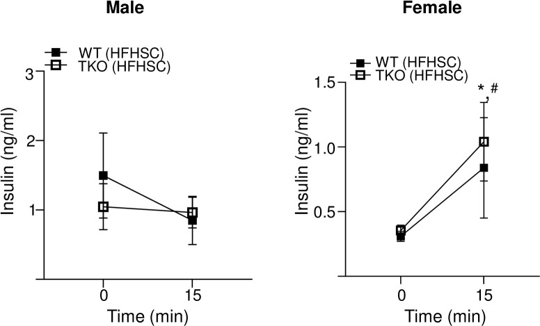
Glucose stimulated insulin secretion (GSIS) was assessed in WT and TKO mice fed HFHSC diet for 14 weeks by determining blood insulin levels before and 15 minutes after glucose injection in mice fasted for 16 h. There was no significant GSIS in male WT and TKO mice fed the HFHSC diet indicating diet-induced β-cell dysfunction (Fig 6, left panel). Both WT and TKO female mice showed significant increase in insulin secretion in response to glucose stimulation and there was no significant difference between the two strains (Fig 6, right panel).

Fig 6. Deficiency of SAA does not affect glucose stimulated insulin secretion.

Fig 6

Plasma insulin levels in male (left panel) and female (right panel) mice before and 15 minutes after intraperitoneal glucose injection, the mice were fed HFHSC diet for 14 weeks and fasted for 16 h prior to the study. Data are mean ±SEM;. * and # = P ≤ 0.05 between * WT chow and HFHSC diet and # between TKO chow and HFHSC diet.

Deficiency of SAA does not impact diet-induced dyslipidemia or hepatic lipogenic gene expression

Earlier studies by Ather and Poynter [44] demonstrated that lack of SAA3 in mice promoted adult onset weight gain, hepatosteatosis and dyslipidemia in addition to significant weight gain with high-fat diet compared to WT litter controls. However, den Hartigh et al. [28] demonstrated that SAA3-deficient mice lost weight in response to an obesogenic diet and female SAA3-deficient mice but not male mice developed improved lipoprotein profiles and plasma lipid levels compared to WT litter controls. We investigated whether deficiency of SAA impacts plasma or liver lipid levels or hepatic lipogenic gene expression with or without HFHSC diet challenge. Fasting plasma triglyceride (TG) and plasma non-esterified fatty acids (NEFA) levels were similar for all groups of mice (S3A and S3B Fig). Plasma total cholesterol (TC) increased significantly with HFHSC diet in both WT and TKO mice, however there were no significant difference in TC between the two strains of mice (S3C Fig). Plasma HDL levels increased significantly with HFHSC diet in WT and TKO female but not male mice (S3D Fig). However, there were no significant differences in HDL between the two strains of male or female mice with either HFHSC or chow diet (S3D Fig). Liver TG increased significantly following HFHSC diet in both the strains of mice (S4A Fig), but there were no significant strain-dependent differences in liver TG with either chow or HFHSC diet (S4A Fig). Liver TC increased significantly with HFHSC diet only in male WT mice (S4B Fig); there were no significant changes in liver TC between any other groups of mice compared (S4B Fig). Hepatic lipogenic gene expression was analyzed in WT and TKO mice fed either chow or HFHSC diet for 16 weeks. There were no significant strain-specific differences in SREBP-1c, SCD-1, HMGCoAR or FAS mRNA abundance in livers of mice fed either chow or HFHSC diet (S5A–S5D Fig).

Deletion of SAA does not affect adipose tissue inflammation in mice fed HFHSC diet

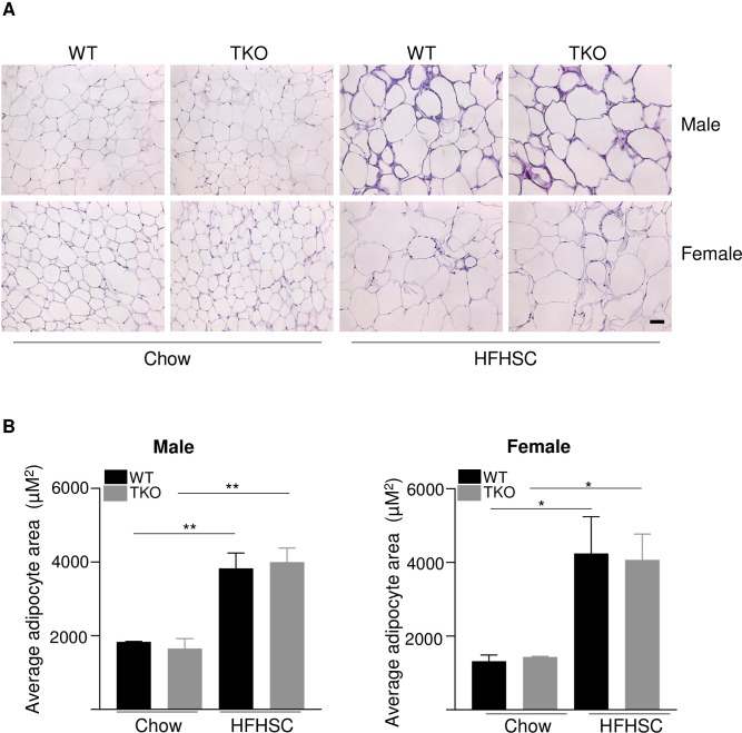
Adipose tissue inflammation and immune cell recruitment are hallmarks of obesity and play critical roles in the development of insulin resistance and glucose intolerance. Histological staining of gonadal fat sections from chow and HFHSC diet-fed WT and TKO mice showed no apparent differences in morphology between the two strains of mice. While there was a significant increase in adipocyte size with HFHSC diet feeding in both male and female mice. No significant strain-dependent differences were observed in female mice with either diet (Fig 7A and 7B).

Fig 7. Deficiency of SAA does not affect diet-induced changes in adipose tissue structure.

Fig 7

A) Representative images of HE staining in gonadal white adipose tissues in male and female WT and TKO mice fed either chow or HFHSC diet for 16 weeks. Scale bar 50 μm. B) adipocyte area was determined for 3 randomly chosen sections for 3 mice/strain; male (left) and female (right) mice; data are mean ±SEM;. * and ** = P ≤ 0.05 and ≤ 0.01 respectively.

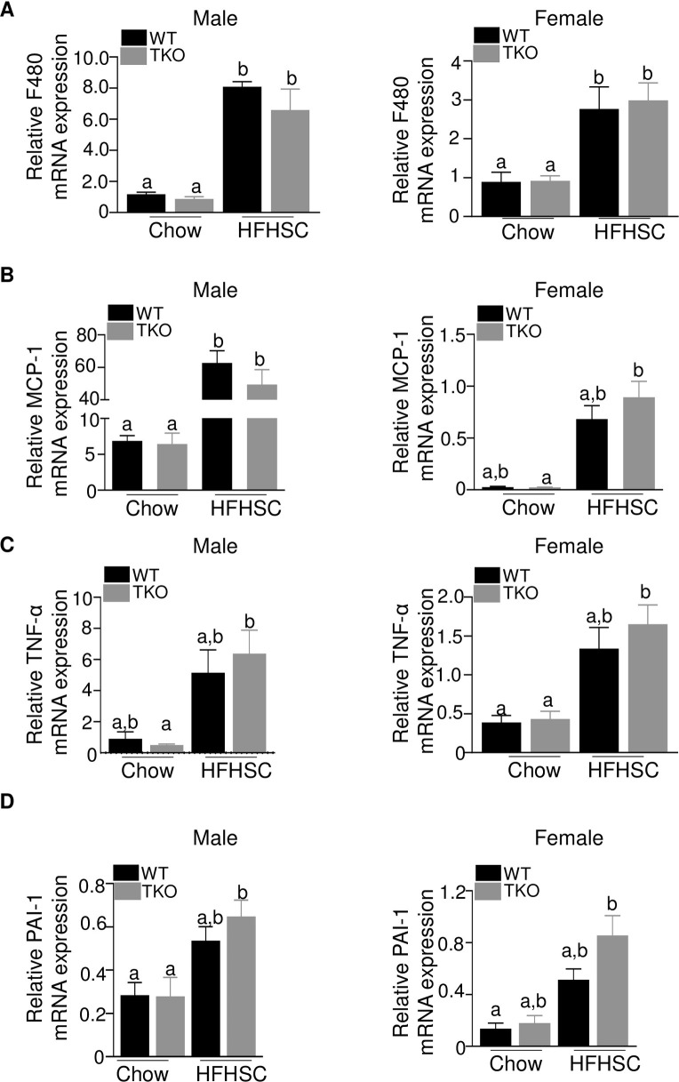
Male and female mice displayed increased amounts of F4/80 mRNA in gonadal adipose tissue in response to HFHSC diet feeding (Fig 8A), indicating increased macrophage content in the obese mice. MCP-1, a chemokine implicated in the recruitment of inflammatory cells in obesity [45], was also increased in adipose tissue of mice fed HFHSC diet (Fig 8B). Moreover, F4/80 and MCP-1 mRNA abundance was similar in adipose tissue of WT and TKO mice fed the obesogenic diet indicating a lack of effect of SAA on obesity-associated adipose tissue inflammation (Fig 8A and 8B). Other indices of adipose tissue inflammation, including TNF-α (Fig 8C), PAI-1 (Fig 8D) were not significantly altered by SAA deficiency in either male or female mice after obesogenic diet. IL-6 or IL-1β mRNA levels did not change significantly irrespective of diet, gender or strain (S6A and S6B Fig). Plasma endotoxin levels were comparable among the different groups of mice (S6C Fig).

Fig 8. Deficiency of SAA does not affect diet-induced changes in adipose tissue inflammatory status.

Fig 8

A-D) Expression of F480, MCP-1, TNFα and PAI-1genes respectively in the gonadal adipose tissues of male and female WT and TKO mice fed either chow or HFHSC diet for 16 weeks (n = 4-10/group). Data are mean ±SEM; data that are not significantly different (P>0.05) are indicated with the same letter.

Discussion

In this study, using male and female SAA-deficient mouse model (deficient in all three inducible SAA isoforms), we investigated if SAA has any role in the development of diet-induced obesity, adipose tissue inflammation, and impairment in glucose or lipid metabolism. The major finding from the study is that while SAA levels in adipose tissue, liver and in circulation are markedly increased with obesity, deficiency of SAA does not impact the development of obesity or obesogenic diet-induced adipose tissue inflammation. Contrary to the thought that SAA may promote obesity related impairment in glucose homeostasis, the present study shows a modest worsening of glucose tolerance in SAA-deficient mice (TKO) compared to the WT mice on HFHSC diet. However, there was no significant effects on insulin tolerance or GSIS. SAA does not contribute to diet-induced dyslipidemia or altered liver lipid homeostasis. Thus, taken together, our study confirms that in mice, the expression of inducible SAA isoforms is significantly increased in liver and adipose tissues after obesogenic diet feeding. However, this induction appears to be a consequence, not a cause, of diet-induced obesity and adipose tissue inflammation.

A number of studies in the past have shown altered SAA levels in circulation and in adipose tissues with changes in diet and body weight, raising the possibility that SAA plays a role in the development of obesity and/or obesity-related complications [1518]. Studies by Sjoholm and colleagues suggest a sexual dimorphic role of SAA in obesity and obesity-associated inflammation [46]. In vivo studies have provided contradictory reports on the roles of SAA in obesity and associated metabolic complications. For example, some authors have reported that SAA3 suppresses adult onset and diet-induced weight gain. However, contrary to this conclusion, other authors have reported that SAA3 exacerbates weight gain induced by an obesogenic diet in a sexually dimorphic manner [28]. Vercalsteren et al. reported that SAA3 gene silencing impaired adipogenesis by studies in vitro in murine preadipocyte cell lines (3T3-F442A), when SAA3-silenced preadipocytes were implanted into BALB/c Nude mice, the mice developed smaller fat pads than their control counterparts [47]. Silencing SAA1.1 and SAA2.1 expression in Swiss Webster mice by antisense oligonucleotides was reported to reduce adipose tissue expansion significantly in these mice along with suppressed adipose tissue inflammation and improved glucose and insulin tolerance in mice fed high fat diet [30]. Differences in body weight or body fat gain between WT and TKO mice fed either chow or HFHSC diet were not observed in the current study (Fig 3A–3C). Phenotypic differences observed between this study and the earlier studies [28, 44, 47] could be either due the differences in mouse strains used, mice with deficiency of all three SAA isoforms vs mice with SAA3 or SAA1.1/2.1 deficiency or the diet used. SAA3 is considered a pseudogene in humans [7] hence the results from the studies on SAA3 have no relevance to humans. However, the presence of SAA3 cannot be ignored in animal studies due its possible biological effects that may or may not overlap with SAA1.1 and SAA2.1 [25]. To our knowledge this is the only study where mice deficient in all the three inducible isoforms of SAA were used to investigate the role of SAA in obesity and associated metabolic complications.

In vitro studies have also implicated SAA in obesity and obesity-associated metabolic perturbations. Incubating 3T3-L1 preadipocytes with recombinant hSAA1 was shown to result in enhanced proliferation, decreased differentiation and altered insulin sensitivity [48]. Results from several in vitro studies could not be reproduced when tested in vivo. For example, a number of in vitro studies show SAA to have pro-inflammatory properties (reviewed in [7, 49]), however, such activities were not observed with manipulation of SAA expression in vivo [21, 50]. Several in vitro studies have used commercially available recombinant SAA which has two amino acid substitutions (at positions 61 and 71) compared to native and thus may show activities different from either mouse or human SAA [51, 52]. Discrepancies in properties between recombinant SAA and endogenous SAA purified from acute-phase plasma have been shown in vitro [25]. Possible lipopolysaccharide contamination of E. coli-derived recombinant SAA could contribute to some of the proinflammatory activities exhibited by this protein [53]. Hence results from all papers published using recombinant SAA for in vitro studies should be reinterpreted with caution. Furthermore, it is now recognized that activities observed for lipid-free SAA in vitro are lost when SAA is lipid-associated, the form SAA is believed to exist in vivo [21, 26, 54].

The existence of multiple isoforms also poses a challenge to SAA studies. Potential functional differences between the three murine acute phase SAA isoforms have not be comprehensively examined. Though mouse SAA1.1 and 2.1 share 91% protein sequence identity [25], differences in properties of the two isoforms have been observed in its amyloidogenic potential [55]. Mouse SAA3 shares ~67% sequence identity to mouse SAA1.1 and SAA2.1 [25], yet differs from the other two with regard to its expression profile and HDL association [8]. Further, there exists a less studied minor acute-phase [56] or a constitutive [11, 57] isoform of SAA, SAA4, which contributes to more than 90% of total circulating SAAs in the absence of inflammation [58]. Mouse models deficient in all four isoforms of SAA including SAA4 have been developed recently and studied in the context of retinol-binding and adaptive immunity [5961]. The contributions of SAA4 if any, to obesity and glucose homeostasis need to be investigated in future. Our data indicate that deletion of all inducible SAA isoforms, including SAA3, does not impact weight gain, adipose tissue inflammation, or major metabolic consequences that occur in mice with obesogenic diet feeding. Whether deletion of individual isoforms of SAA produce different effects cannot be ruled out at this time and is a limitation of our study.

Based on human population studies, elevated levels of circulating SAA have been recognized as a risk factor for certain chronic inflammatory diseases for many decades, including atherosclerotic cardiovascular disease [49] and cancer [62]. The development of mice with targeted deletion of the inducible SAAs allowed us to identify a causative role for SAA in both atherosclerosis [32, 36] and metastatic pancreatic cancer. Here we report that the loss of all three inducible SAAs has no effect on the development of diet-induced obesity or adipose tissue inflammation, with a modest but significant protective effect on glucose intolerance. Thus, increased SAA in the setting of obesity appears to be a consequence, not a cause, of adipose tissue inflammation.

Supporting information

S1 Fig

A) Genotyping to identify SAA TKO mice are done by a multi-step process. The first step is genotyping for SAA1/2 KO utilizing a 3 primer PCR reaction as described earlier (de Beer et al., J Lipid Res. 2010. 51:3117–3125). The genotype to determine SAA 3 deficiency is performed by two sets of PCRs, using forward primer for the PCRs designed to detect WT sequence or SAA3 KO (CRISPR-Cas9) sequences respectively. The primers for the two PCRs are shown on the right panel. The generation of TKO mice from SAA1.1/SAA2.1-deficient mice is described earlier [35]. B) Expression of SAA3 mRNA in the adipose tissues of male (left panel) and female (right panel) WT and TKO mice fed either chow or HFHSC diet for 16 weeks. C) SAA4 mRNA expression in the adipose tissues of male (left panel) and female (right panel) WT and TKO mice fed either chow or HFHSC diet for 16 weeks. D) Expression of SAA3 mRNA in the livers of male (left panel) and female (right panel) WT and TKO mice fed either chow or HFHSC diet for 16 weeks. Data are mean ±SEM; ** = P ≤ 0.01 and *** = P ≤ 0.001.

(PPTX)

S2 Fig. Deficiency of SAA does not affect basal insulin tolerance in mice.

Insulin tolerance test (ITT) was performed in male (left panel) and female (right panel) WT and TKO mice on chow diet (n = 4–5 mice/group).

(PPTX)

S3 Fig. Deficiency of SAA does not significantly impact diet-induced dyslipidemia.

Plasma triglycerides A), non-esterified fatty acids B) and total cholesterol C) levels in male (left panel) and female (right panel) WT and TKO mice (n = 4–15) fed either chow or HFHSC diet for 16 weeks. D) Plasma HDL levels in male (left panel) and female (right panel) WT and TKO mice (n = 4–15) fed either chow or HFHSC diet for 16 weeks. Data are mean ±SEM; data that are not significantly different (P>0.05) are indicated with the same letter.

(PPTX)

S4 Fig. Deficiency of SAA does not significantly impact diet-induced changes in hepatic triglycerides or cholesterol levels.

Liver triglycerides A) and total cholesterol B) levels in male (left panel) and female (right panel) WT and TKO mice (n = 5–15) fed either chow or HFHSC diet for 16 weeks. Data are mean ±SEM; data that are not significantly different (P>0.05) are indicated with the same letter.

(PPTX)

S5 Fig. Deficiency of SAA does not significantly impact diet-induced changes in hepatic lipogenic gene expression.

Expression of SREBP1-c, SCD-1, HMGCoAR and FAS genes (A-D) respectively in the livers of male (left panel) and female (right panel) WT and TKO mice fed either chow or HFHSC diet for 16 weeks (n = 4-10/group). Data are mean ±SEM; data that are not significantly different (P>0.05) are indicated with the same letter.

(PPTX)

S6 Fig. Deficiency of SAA does not significantly impact diet-induced changes in adipose tissue IL-6 and IL-1β gene expression levels.

Expression of IL-6 A) and IL-1β B) mRNA in the adipose tissues of male (left panel) and female (right panel) WT and TKO mice fed either chow or HFHSC diet for 16 weeks (n = 4-10/group). C) Plasma endotoxin levels at the study termination in male (left panel) and female (right panel) WT and TKO mice fed either chow or HFHSC diet for 16 weeks (n = 4-10/group). Data are mean ±SEM.

(PPTX)

S1 Raw images

(PDF)

S2 Raw images

(PDF)

S1 Data

(XLSX)

Data Availability

All relevant data are within the paper and its Supporting Information files.

Funding Statement

This study was funded by the Diabetes Research Center at Washington University in St. Louis of the National Institutes of Health in the form of a grant (No. P30DK020579) to PS. This study was also funded by the National Institutes of Health Grants in the form of grants to PS and LT (No. HL147381), and NW and FD (No. HL134731). This study was also funded by the Department of Veterans Affairs in the form of a grant to LT (No. BX004275). This study was also supported with resources and facilities provided by the Centers of Biomedical Research Excellence (COBRE) at the University of Kentucky, which was supported by an Institutional Development Award (IDeA) from the National Institute of General Medical Sciences of the National Institutes of Health under a grant (No. P30 GM127211). The funders had no role in study design, data collection, and analysis, decision to publish, or preparation of the manuscript.

References

  • 1.Hotamisligil GS. Inflammation and metabolic disorders. Nature. 2006;444(7121):860–7. 10.1038/nature05485 . [DOI] [PubMed] [Google Scholar]
  • 2.McNelis JC, Olefsky JM. Macrophages, immunity, and metabolic disease. Immunity. 2014;41(1):36–48. 10.1016/j.immuni.2014.05.010 . [DOI] [PubMed] [Google Scholar]
  • 3.Osborn O, Olefsky JM. The cellular and signaling networks linking the immune system and metabolism in disease. Nat Med. 2012;18(3):363–74. 10.1038/nm.2627 . [DOI] [PubMed] [Google Scholar]
  • 4.Yamamoto K, Goto N, Kosaka J, Shiroo M, Yeul YD, Migita S. Structural diversity of murine serum amyloid A genes. Evolutionary implications. J Immunol. 1987;139(5):1683–8. . [PubMed] [Google Scholar]
  • 5.Webb NR. High-Density Lipoproteins and Serum Amyloid A (SAA). Curr Atheroscler Rep. 2021;23(2):7. 10.1007/s11883-020-00901-4 . [DOI] [PMC free article] [PubMed] [Google Scholar]
  • 6.Kluve-Beckerman B, Drumm ML, Benson MD. Nonexpression of the human serum amyloid A three (SAA3) gene. DNA Cell Biol. 1991;10(9):651–61. 10.1089/dna.1991.10.651 [DOI] [PubMed] [Google Scholar]
  • 7.Sack GH Jr. Serum amyloid A—a review. Mol Med. 2018;24(1):46. 10.1186/s10020-018-0047-0 . [DOI] [PMC free article] [PubMed] [Google Scholar]
  • 8.Tannock LR, De Beer MC, Ji A, Shridas P, Noffsinger VP, den Hartigh L, et al. Serum amyloid A3 is a high density lipoprotein-associated acute-phase protein. J Lipid Res. 2018;59(2):339–47. 10.1194/jlr.M080887 . [DOI] [PMC free article] [PubMed] [Google Scholar]
  • 9.Calabro P, Riegler L, Maddaloni V, Fimiani F, Limongelli G, Martone F, et al. Adipose tissue as an endocrine organ: production of serum amyloid a in response to inflammatory cytokines by human adipocytes. European Heart Journal. 2013;34(suppl_1). 10.1093/eurheartj/eht309.P4160 [DOI] [Google Scholar]
  • 10.Poitou C, Coussieu C, Rouault C, Coupaye M, Cancello R, Bedel JF, et al. Serum amyloid A: a marker of adiposity-induced low-grade inflammation but not of metabolic status. Obesity (Silver Spring). 2006;14(2):309–18. 10.1038/oby.2006.40 . [DOI] [PubMed] [Google Scholar]
  • 11.Whitehead AS, de Beer MC, Steel DM, Rits M, Lelias JM, Lane WS, et al. Identification of novel members of the serum amyloid A protein superfamily as constitutive apolipoproteins of high density lipoprotein. J Biol Chem. 1992;267(6):3862–7. . [PubMed] [Google Scholar]
  • 12.Jahangiri A, Wilson PG, Hou T, Brown A, King VL, Tannock LR. Serum amyloid A is found on ApoB-containing lipoproteins in obese humans with diabetes. Obesity (Silver Spring). 2013;21(5):993–6. 10.1002/oby.20126 . [DOI] [PMC free article] [PubMed] [Google Scholar]
  • 13.van Dielen FM, van’t Veer C, Schols AM, Soeters PB, Buurman WA, Greve JW. Increased leptin concentrations correlate with increased concentrations of inflammatory markers in morbidly obese individuals. Int J Obes Relat Metab Disord. 2001;25(12):1759–66. 10.1038/sj.ijo.0801825 . [DOI] [PubMed] [Google Scholar]
  • 14.Yang RZ, Lee MJ, Hu H, Pollin TI, Ryan AS, Nicklas BJ, et al. Acute-phase serum amyloid A: an inflammatory adipokine and potential link between obesity and its metabolic complications. PLoS Med. 2006;3(6):e287. 10.1371/journal.pmed.0030287 . [DOI] [PMC free article] [PubMed] [Google Scholar]
  • 15.Gomez-Ambrosi J, Salvador J, Rotellar F, Silva C, Catalan V, Rodriguez A, et al. Increased serum amyloid A concentrations in morbid obesity decrease after gastric bypass. Obes Surg. 2006;16(3):262–9. 10.1381/096089206776116525 . [DOI] [PubMed] [Google Scholar]
  • 16.Poitou C, Viguerie N, Cancello R, De Matteis R, Cinti S, Stich V, et al. Serum amyloid A: production by human white adipocyte and regulation by obesity and nutrition. Diabetologia. 2005;48(3):519–28. 10.1007/s00125-004-1654-6 . [DOI] [PubMed] [Google Scholar]
  • 17.Sjoholm K, Palming J, Olofsson LE, Gummesson A, Svensson PA, Lystig TC, et al. A microarray search for genes predominantly expressed in human omental adipocytes: adipose tissue as a major production site of serum amyloid A. J Clin Endocrinol Metab. 2005;90(4):2233–9. 10.1210/jc.2004-1830 . [DOI] [PubMed] [Google Scholar]
  • 18.O’Brien KD, Brehm BJ, Seeley RJ, Bean J, Wener MH, Daniels S, et al. Diet-induced weight loss is associated with decreases in plasma serum amyloid a and C-reactive protein independent of dietary macronutrient composition in obese subjects. J Clin Endocrinol Metab. 2005;90(4):2244–9. 10.1210/jc.2004-1011 . [DOI] [PubMed] [Google Scholar]
  • 19.Marzi C, Huth C, Herder C, Baumert J, Thorand B, Rathmann W, et al. Acute-phase serum amyloid A protein and its implication in the development of type 2 diabetes in the KORA S4/F4 study. Diabetes Care. 2013;36(5):1321–6. 10.2337/dc12-1514 . [DOI] [PMC free article] [PubMed] [Google Scholar]
  • 20.Badolato R, Wang JM, Murphy WJ, Lloyd AR, Michiel DF, Bausserman LL, et al. Serum amyloid A is a chemoattractant: induction of migration, adhesion, and tissue infiltration of monocytes and polymorphonuclear leukocytes. J Exp Med. 1994;180(1):203–9. 10.1084/jem.180.1.203 [DOI] [PMC free article] [PubMed] [Google Scholar]
  • 21.Kim MH, de Beer MC, Wroblewski JM, Webb NR, de Beer FC. SAA does not induce cytokine production in physiological conditions. Cytokine. 2013;61(2):506–12. 10.1016/j.cyto.2012.10.019 . [DOI] [PMC free article] [PubMed] [Google Scholar]
  • 22.Sandri S, Rodriguez D, Gomes E, Monteiro HP, Russo M, Campa A. Is serum amyloid A an endogenous TLR4 agonist? J Leukoc Biol. 2008;83(5):1174–80. 10.1189/jlb.0407203 . [DOI] [PubMed] [Google Scholar]
  • 23.Furlaneto CJ, Campa A. A novel function of serum amyloid A: a potent stimulus for the release of tumor necrosis factor-alpha, interleukin-1beta, and interleukin-8 by human blood neutrophil. Biochem Biophys Res Commun. 2000;268(2):405–8. 10.1006/bbrc.2000.2143 . [DOI] [PubMed] [Google Scholar]
  • 24.Hatanaka E, Pereira Ribeiro F, Campa A. The acute phase protein serum amyloid A primes neutrophils. FEMS Immunol Med Microbiol. 2003;38(1):81–4. 10.1016/S0928-8244(03)00112-3 . [DOI] [PubMed] [Google Scholar]
  • 25.Shridas P, Patrick AC, Tannock LR. Role of Serum Amyloid A in Abdominal Aortic Aneurysm and Related Cardiovascular Diseases. Biomolecules. 2021;11(12). 10.3390/biom11121883 . [DOI] [PMC free article] [PubMed] [Google Scholar]
  • 26.Shridas P, De Beer MC, Webb NR. High-density lipoprotein inhibits serum amyloid A-mediated reactive oxygen species generation and NLRP3 inflammasome activation. J Biol Chem. 2018;293(34):13257–69. 10.1074/jbc.RA118.002428 . [DOI] [PMC free article] [PubMed] [Google Scholar]
  • 27.Eklund KK, Niemi K, Kovanen PT. Immune functions of serum amyloid A. Crit Rev Immunol. 2012;32(4):335–48. 10.1615/critrevimmunol.v32.i4.40 . [DOI] [PubMed] [Google Scholar]
  • 28.den Hartigh LJ, Wang S, Goodspeed L, Ding Y, Averill M, Subramanian S, et al. Deletion of serum amyloid A3 improves high fat high sucrose diet-induced adipose tissue inflammation and hyperlipidemia in female mice. PLoS One. 2014;9(9):e108564. 10.1371/journal.pone.0108564 . [DOI] [PMC free article] [PubMed] [Google Scholar]
  • 29.Ahlin S, Olsson M, Olsson B, Svensson PA, Sjoholm K. No evidence for a role of adipose tissue-derived serum amyloid a in the development of insulin resistance or obesity-related inflammation in hSAA1(+/-) transgenic mice. PLoS One. 2013;8(8):e72204. 10.1371/journal.pone.0072204 . [DOI] [PMC free article] [PubMed] [Google Scholar]
  • 30.de Oliveira EM, Ascar TP, Silva JC, Sandri S, Migliorini S, Fock RA, et al. Serum amyloid A links endotoxaemia to weight gain and insulin resistance in mice. Diabetologia. 2016;59(8):1760–8. 10.1007/s00125-016-3970-z . [DOI] [PubMed] [Google Scholar]
  • 31.Dong Z, Wu T, Qin W, An C, Wang Z, Zhang M, et al. Serum amyloid A directly accelerates the progression of atherosclerosis in apolipoprotein E-deficient mice. Mol Med. 2011;17(11–12):1357–64. 10.2119/molmed.2011.00186 . [DOI] [PMC free article] [PubMed] [Google Scholar]
  • 32.Thompson JC, Jayne C, Thompson J, Wilson PG, Yoder MH, Webb N, et al. A brief elevation of serum amyloid A is sufficient to increase atherosclerosis. J Lipid Res. 2015;56(2):286–93. 10.1194/jlr.M054015 . [DOI] [PMC free article] [PubMed] [Google Scholar]
  • 33.Webb NR, De Beer MC, Wroblewski JM, Ji A, Bailey W, Shridas P, et al. Deficiency of Endogenous Acute-Phase Serum Amyloid A Protects apoE-/- Mice From Angiotensin II-Induced Abdominal Aortic Aneurysm Formation. Arterioscler Thromb Vasc Biol. 2015;35(5):1156–65. 10.1161/ATVBAHA.114.304776 . [DOI] [PMC free article] [PubMed] [Google Scholar]
  • 34.King VL, Hatch NW, Chan HW, de Beer MC, de Beer FC, Tannock LR. A murine model of obesity with accelerated atherosclerosis. Obesity (Silver Spring). 2010;18(1):35–41. 10.1038/oby.2009.176 . [DOI] [PMC free article] [PubMed] [Google Scholar]
  • 35.Lee JY, Hall JA, Kroehling L, Wu L, Najar T, Nguyen HH, et al. Serum Amyloid A Proteins Induce Pathogenic Th17 Cells and Promote Inflammatory Disease. Cell. 2020;180(1):79–91 e16. 10.1016/j.cell.2019.11.026 . [DOI] [PMC free article] [PubMed] [Google Scholar]
  • 36.Thompson JC, Wilson PG, Shridas P, Ji A, de Beer M, de Beer FC, et al. Serum amyloid A3 is pro-atherogenic. Atherosclerosis. 2018;268:32–5. 10.1016/j.atherosclerosis.2017.11.011 . [DOI] [PMC free article] [PubMed] [Google Scholar]
  • 37.de Beer MC, Webb NR, Wroblewski JM, Noffsinger VP, Rateri DL, Ji A, et al. Impact of serum amyloid A on high density lipoprotein composition and levels. J Lipid Res. 2010;51(11):3117–25. 10.1194/jlr.M005413 . [DOI] [PMC free article] [PubMed] [Google Scholar]
  • 38.Subramanian S, Han CY, Chiba T, McMillen TS, Wang SA, Haw A 3rd, et al. Dietary cholesterol worsens adipose tissue macrophage accumulation and atherosclerosis in obese LDL receptor-deficient mice. Arterioscler Thromb Vasc Biol. 2008;28(4):685–91. 10.1161/ATVBAHA.107.157685 . [DOI] [PMC free article] [PubMed] [Google Scholar]
  • 39.Shridas P, Zahoor L, Forrest KJ, Layne JD, Webb NR. Group X secretory phospholipase A2 regulates insulin secretion through a cyclooxygenase-2-dependent mechanism. J Biol Chem. 2014;289(40):27410–7. 10.1074/jbc.M114.591735 . [DOI] [PMC free article] [PubMed] [Google Scholar]
  • 40.Softic S, Gupta MK, Wang GX, Fujisaka S, O’Neill BT, Rao TN, et al. Divergent effects of glucose and fructose on hepatic lipogenesis and insulin signaling. J Clin Invest. 2018;128(3):1199. 10.1172/JCI99009 . [DOI] [PMC free article] [PubMed] [Google Scholar]
  • 41.Folch J, Lees M, Sloane Stanley GH. A simple method for the isolation and purification of total lipides from animal tissues. J Biol Chem. 1957;226(1):497–509. . [PubMed] [Google Scholar]
  • 42.Chiba T, Han CY, Vaisar T, Shimokado K, Kargi A, Chen MH, et al. Serum amyloid A3 does not contribute to circulating SAA levels. J Lipid Res. 2009;50(7):1353–62. 10.1194/jlr.M900089-JLR200 . [DOI] [PMC free article] [PubMed] [Google Scholar]
  • 43.De Buck M, Gouwy M, Wang JM, Van Snick J, Opdenakker G, Struyf S, et al. Structure and Expression of Different Serum Amyloid A (SAA) Variants and their Concentration-Dependent Functions During Host Insults. Curr Med Chem. 2016;23(17):1725–55. 10.2174/0929867323666160418114600 . [DOI] [PMC free article] [PubMed] [Google Scholar]
  • 44.Ather JL, Poynter ME. Serum amyloid A3 is required for normal weight and immunometabolic function in mice. PLoS One. 2018;13(2):e0192352. 10.1371/journal.pone.0192352 . [DOI] [PMC free article] [PubMed] [Google Scholar]
  • 45.Kanda H, Tateya S, Tamori Y, Kotani K, Hiasa K, Kitazawa R, et al. MCP-1 contributes to macrophage infiltration into adipose tissue, insulin resistance, and hepatic steatosis in obesity. J Clin Invest. 2006;116(6):1494–505. 10.1172/JCI26498 . [DOI] [PMC free article] [PubMed] [Google Scholar]
  • 46.Sjoholm K, Lundgren M, Olsson M, Eriksson JW. Association of serum amyloid A levels with adipocyte size and serum levels of adipokines: differences between men and women. Cytokine. 2009;48(3):260–6. 10.1016/j.cyto.2009.08.005 . [DOI] [PubMed] [Google Scholar]
  • 47.Vercalsteren E, Vranckx C, Vermeire I, Gooijen M, Lijnen R, Scroyen I. Serum amyloid A3 deficiency impairs in vitro and in vivo adipocyte differentiation. Adipocyte. 2021;10(1):242–50. 10.1080/21623945.2021.1916220 . [DOI] [PMC free article] [PubMed] [Google Scholar]
  • 48.Filippin-Monteiro FB, de Oliveira EM, Sandri S, Knebel FH, Albuquerque RC, Campa A. Serum amyloid A is a growth factor for 3T3-L1 adipocytes, inhibits differentiation and promotes insulin resistance. Int J Obes (Lond). 2012;36(8):1032–9. 10.1038/ijo.2011.193 . [DOI] [PMC free article] [PubMed] [Google Scholar]
  • 49.Shridas P, Tannock LR. Role of serum amyloid A in atherosclerosis. Curr Opin Lipidol. 2019;30(4):320–5. 10.1097/MOL.0000000000000616 . [DOI] [PMC free article] [PubMed] [Google Scholar]
  • 50.Simons JP, Al-Shawi R, Ellmerich S, Speck I, Aslam S, Hutchinson WL, et al. Pathogenetic mechanisms of amyloid A amyloidosis. Proc Natl Acad Sci U S A. 2013;110(40):16115–20. doi: 10.1073/pnas.1306621110 . [DOI] [PMC free article] [PubMed] [Google Scholar]
  • 51.Bjorkman L, Raynes JG, Shah C, Karlsson A, Dahlgren C, Bylund J. The proinflammatory activity of recombinant serum amyloid A is not shared by the endogenous protein in the circulation. Arthritis Rheum. 2010;62(6):1660–5. 10.1002/art.27440 . [DOI] [PubMed] [Google Scholar]
  • 52.Christenson K, Bjorkman L, Ahlin S, Olsson M, Sjoholm K, Karlsson A, et al. Endogenous Acute Phase Serum Amyloid A Lacks Pro-Inflammatory Activity, Contrasting the Two Recombinant Variants That Activate Human Neutrophils through Different Receptors. Front Immunol. 2013;4:92. 10.3389/fimmu.2013.00092 . [DOI] [PMC free article] [PubMed] [Google Scholar]
  • 53.Abouelasrar Salama S, Gouwy M, Van Damme J, Struyf S. The turning away of serum amyloid A biological activities and receptor usage. Immunology. 2021;163(2):115–27. 10.1111/imm.13295 . [DOI] [PMC free article] [PubMed] [Google Scholar]
  • 54.Franco AG, Sandri S, Campa A. High-density lipoprotein prevents SAA-induced production of TNF-alpha in THP-1 monocytic cells and peripheral blood mononuclear cells. Mem Inst Oswaldo Cruz. 2011;106(8):986–92. 10.1590/s0074-02762011000800014 . [DOI] [PubMed] [Google Scholar]
  • 55.Yu J, Zhu H, Guo JT, de Beer FC, Kindy MS. Expression of mouse apolipoprotein SAA1.1 in CE/J mice: isoform-specific effects on amyloidogenesis. Lab Invest. 2000;80(12):1797–806. 10.1038/labinvest.3780191 . [DOI] [PubMed] [Google Scholar]
  • 56.Yamada T, Miyake N, Itoh K, Igari J. Further characterization of serum amyloid A4 as a minor acute phase reactant and a possible nutritional marker. Clin Chem Lab Med. 2001;39(1):7–10. 10.1515/CCLM.2001.003 . [DOI] [PubMed] [Google Scholar]
  • 57.Steel DM, Donoghue FC, O’Neill RM, Uhlar CM, Whitehead AS. Expression and regulation of constitutive and acute phase serum amyloid A mRNAs in hepatic and non-hepatic cell lines. Scand J Immunol. 1996;44(5):493–500. 10.1046/j.1365-3083.1996.d01-341.x . [DOI] [PubMed] [Google Scholar]
  • 58.de Beer MC, Yuan T, Kindy MS, Asztalos BF, Roheim PS, de Beer FC. Characterization of constitutive human serum amyloid A protein (SAA4) as an apolipoprotein. J Lipid Res. 1995;36(3):526–34. . [PubMed] [Google Scholar]
  • 59.Bang YJ, Hu Z, Li Y, Gattu S, Ruhn KA, Raj P, et al. Serum amyloid A delivers retinol to intestinal myeloid cells to promote adaptive immunity. Science. 2021;373(6561):eabf9232. 10.1126/science.abf9232 . [DOI] [PMC free article] [PubMed] [Google Scholar]
  • 60.Derebe MG, Zlatkov CM, Gattu S, Ruhn KA, Vaishnava S, Diehl GE, et al. Serum amyloid A is a retinol binding protein that transports retinol during bacterial infection. Elife. 2014;3:e03206. 10.7554/eLife.03206 . [DOI] [PMC free article] [PubMed] [Google Scholar]
  • 61.Hu Z, Bang YJ, Ruhn KA, Hooper LV. Molecular basis for retinol binding by serum amyloid A during infection. Proc Natl Acad Sci U S A. 2019;116(38):19077–82. 10.1073/pnas.1910713116 . [DOI] [PMC free article] [PubMed] [Google Scholar]
  • 62.Zhou J, Sheng J, Fan Y, Zhu X, Tao Q, He Y, et al. Association between serum amyloid A levels and cancers: a systematic review and meta-analysis. Postgrad Med J. 2018;94(1115):499–507. 10.1136/postgradmedj-2018-136004 . [DOI] [PubMed] [Google Scholar]

Decision Letter 0

Michael Bader

15 Nov 2021

PONE-D-21-32072Serum Amyloid A is not obligatory for high-fat, high-sucrose, cholesterol-fed diet-induced obesity and its metabolic and inflammatory complicationsPLOS ONE

Dear Dr. Shridas,

Thank you for submitting your manuscript to PLOS ONE. After careful consideration, we feel that it has merit but does not fully meet PLOS ONE’s publication criteria as it currently stands. Therefore, we invite you to submit a revised version of the manuscript that addresses the points raised during the review process.

Please submit your revised manuscript by Dec 30 2021 11:59PM. If you will need more time than this to complete your revisions, please reply to this message or contact the journal office at plosone@plos.org. When you're ready to submit your revision, log on to https://www.editorialmanager.com/pone/ and select the 'Submissions Needing Revision' folder to locate your manuscript file.

Please include the following items when submitting your revised manuscript:

  • A rebuttal letter that responds to each point raised by the academic editor and reviewer(s). You should upload this letter as a separate file labeled 'Response to Reviewers'.

  • A marked-up copy of your manuscript that highlights changes made to the original version. You should upload this as a separate file labeled 'Revised Manuscript with Track Changes'.

  • An unmarked version of your revised paper without tracked changes. You should upload this as a separate file labeled 'Manuscript'.

If you would like to make changes to your financial disclosure, please include your updated statement in your cover letter. Guidelines for resubmitting your figure files are available below the reviewer comments at the end of this letter.

If applicable, we recommend that you deposit your laboratory protocols in protocols.io to enhance the reproducibility of your results. Protocols.io assigns your protocol its own identifier (DOI) so that it can be cited independently in the future. For instructions see: https://journals.plos.org/plosone/s/submission-guidelines#loc-laboratory-protocols. Additionally, PLOS ONE offers an option for publishing peer-reviewed Lab Protocol articles, which describe protocols hosted on protocols.io. Read more information on sharing protocols at https://plos.org/protocols?utm_medium=editorial-email&utm_source=authorletters&utm_campaign=protocols.

We look forward to receiving your revised manuscript.

Kind regards,

Michael Bader

Academic Editor

PLOS ONE

Journal Requirements:

When submitting your revision, we need you to address these additional requirements.

1. Please ensure that your manuscript meets PLOS ONE's style requirements, including those for file naming. The PLOS ONE style templates can be found at 

https://journals.plos.org/plosone/s/file?id=wjVg/PLOSOne_formatting_sample_main_body.pdf and 

https://journals.plos.org/plosone/s/file?id=ba62/PLOSOne_formatting_sample_title_authors_affiliations.pdf

2. We noticed you have some minor occurrence of overlapping text with the following previous publication(s), which needs to be addressed:

- https://onlinelibrary.wiley.com/doi/10.1002/oby.20126

- https://link.springer.com/article/10.1007%2Fs11883-020-00901-4

The text that needs to be addressed involves the introduction

In your revision ensure you cite all your sources (including your own works), and quote or rephrase any duplicated text outside the methods section. Further consideration is dependent on these concerns being addressed.

3. We note that the grant information you provided in the ‘Funding Information’ and ‘Financial Disclosure’ sections do not match. 

When you resubmit, please ensure that you provide the correct grant numbers for the awards you received for your study in the ‘Funding Information’ section

4. In your Data Availability statement, you have not specified where the minimal data set underlying the results described in your manuscript can be found. PLOS defines a study's minimal data set as the underlying data used to reach the conclusions drawn in the manuscript and any additional data required to replicate the reported study findings in their entirety. All PLOS journals require that the minimal data set be made fully available. For more information about our data policy, please see http://journals.plos.org/plosone/s/data-availability.

"Upon re-submitting your revised manuscript, please upload your study’s minimal underlying data set as either Supporting Information files or to a stable, public repository and include the relevant URLs, DOIs, or accession numbers within your revised cover letter. For a list of acceptable repositories, please see http://journals.plos.org/plosone/s/data-availability#loc-recommended-repositories. Any potentially identifying patient information must be fully anonymized.

Important: If there are ethical or legal restrictions to sharing your data publicly, please explain these restrictions in detail. Please see our guidelines for more information on what we consider unacceptable restrictions to publicly sharing data: http://journals.plos.org/plosone/s/data-availability#loc-unacceptable-data-access-restrictions. Note that it is not acceptable for the authors to be the sole named individuals responsible for ensuring data access.

We will update your Data Availability statement to reflect the information you provide in your cover letter.

5. We note that you have included the phrase “data not shown” in your manuscript. Unfortunately, this does not meet our data sharing requirements. PLOS does not permit references to inaccessible data. We require that authors provide all relevant data within the paper, Supporting Information files, or in an acceptable, public repository. Please add a citation to support this phrase or upload the data that corresponds with these findings to a stable repository (such as Figshare or Dryad) and provide and URLs, DOIs, or accession numbers that may be used to access these data. Or, if the data are not a core part of the research being presented in your study, we ask that you remove the phrase that refers to these data.

[Note: HTML markup is below. Please do not edit.]

Reviewers' comments:

Reviewer's Responses to Questions

Comments to the Author

1. Is the manuscript technically sound, and do the data support the conclusions?

The manuscript must describe a technically sound piece of scientific research with data that supports the conclusions. Experiments must have been conducted rigorously, with appropriate controls, replication, and sample sizes. The conclusions must be drawn appropriately based on the data presented.

Reviewer #1: Yes

Reviewer #2: Yes

Reviewer #3: Yes

**********

2. Has the statistical analysis been performed appropriately and rigorously?

Reviewer #1: Yes

Reviewer #2: Yes

Reviewer #3: Yes

**********

3. Have the authors made all data underlying the findings in their manuscript fully available?

The PLOS Data policy requires authors to make all data underlying the findings described in their manuscript fully available without restriction, with rare exception (please refer to the Data Availability Statement in the manuscript PDF file). The data should be provided as part of the manuscript or its supporting information, or deposited to a public repository. For example, in addition to summary statistics, the data points behind means, medians and variance measures should be available. If there are restrictions on publicly sharing data—e.g. participant privacy or use of data from a third party—those must be specified.

Reviewer #1: Yes

Reviewer #2: Yes

Reviewer #3: Yes

**********

4. Is the manuscript presented in an intelligible fashion and written in standard English?

PLOS ONE does not copyedit accepted manuscripts, so the language in submitted articles must be clear, correct, and unambiguous. Any typographical or grammatical errors should be corrected at revision, so please note any specific errors here.

Reviewer #1: Yes

Reviewer #2: Yes

Reviewer #3: Yes

**********

5. Review Comments to the Author

Please use the space provided to explain your answers to the questions above. You may also include additional comments for the author, including concerns about dual publication, research ethics, or publication ethics. (Please upload your review as an attachment if it exceeds 20,000 characters)

Reviewer #1: The manuscript by Ji et al. explores the contribution of serum amyloid A (SAA) to obesity generation and pathophysiology. They document the diet-induced obesity feeding regimen (high fat, sucrose, and cholesterol) induces SAA expression, but that mice deficient in SAA do not display any substantial or biologically meaningful differences as a consequence of feeding of this diet compared to wildtype mice (an exception perhaps being glucose levels). These results substantiate a lack of involvement of SAAs in diet-induced obesity and associated pathophysiological manifestations. The manuscript is straightforward and the results appropriately presented. However, there are areas of the work that require additional attention in a revised version of the manuscript.

First, critically absent are any data documenting the purported genotype, gene expression absence, and protein abundance decreases in the SAA triple knockout mice. Similar mice have been previously published (PMID 31484771, which are SAA1,2,3,4-deficient), yet the mice used in this manuscript appear to have been generated independently. Consequently, the authors need to present compelling evidence that their mice are what they are claimed to be, and that is mice absent of SAA1, SAA2, and SAA3 proteins. Having never encountered ELISAs that recognize all three SAAs (SAA1/2 are oftentimes both recognized by an SAA ELISA, whereas SAA3 is not unless it is SAA3-specific), several assays will need to be conducted. Furthermore, the aforementioned citation should be included and the new mouse compared to that one.

Second, the authors cited a paper related to the spontaneous development of obesity in SAA3-/- mice fed normal chow (PMID 29390039), but only in the contest of one figure in which it was reported that feeding a HFD for 1 week to the SAA3-/- mice increased weight gain compared to wildtype mice. That was one figure in the paper that otherwise indicated spontaneous obesity in the SAA3-/- mice that was conducted using a different HFD than that used in this manuscript. Additional clarification of these differences should be conveyed by the authors.

Finally, the authors cite papers in which 3T3L1 cells were treated with recombinant SAA, which is problematic due to the fact that recombinant forms of SAA that are generated from E. coli contain contaminants (including TLR2 stimulating lipopeptides). The authors need to mention these findings (PMID) and the citations in that paper that have long suggested a lack of inflammatory activity of SAA proteins. All papers published using recombinant SAA proteins should be reinterpreted with caution, and acknowledgement of the contamination issue needs to be understood by researchers and conveyed by authors in their publications.

Reviewer #2: I think this study adds another piece on the path to elucidating the biological effects of SAA. Some questions have been repeatedly asked: Can the results observed with recombinant SAA be extrapolating to in vivo? Are there free SAA stocks? How much? In serum? in tissues? How do the different forms of SAA work? Is there a regulation between them? How do differences between the biochemistry and metabolism of lipoproteins between species affect the SAA role? Is it possible to extrapolate results from experimental animals to humans? How to establish causal relationships between SAA increase and decrease with some of the effects pointed out for it?

In this complex scenario, the study by Ailing et al. has credit and deserves to be published in PlosOne. The results with the triple deficient in SAA are fair and the experimental design well delineated.

The comparison between males and females is also all interesting.

My comments are:

1- Do the authors have data on food consumption, serum HDL, and endotoxemia comparing TKO animals with WT? If there is any possibility to include these data it will be interesting.

2- It would also have been fantastic to have the data for the single and double deletions, in addition to the triple deletion..... same for super expression.. But I understand perfectly well that this is a mid-term mission.

3- Anyway, I consider that some of these points might be discussed by the authors. Beyond the limitations of previous studies, adding their study limits will provide a clearer picture and put in context the complexity of SAA biology.

Reviewer #3: General comments:

The first sentence of the abstract doesn’t give an appropriate sense of what this manuscript is about, i.e., whether SAA plays a role in the development of obesity and adipose tissue inflammation. It and the second sentence of the abstract should be revised accordingly. The manuscript purports to challenge one proposed role of SAA in obesity, i.e., its role in causing obesity, which is not widely universally accepted based on the limited number of studies. However, the findings in this study show that SAA deletion doesn’t affect the development of obesity and hence doesn’t challenge this proposed role of SAA, which is not well-established. Nor do the finding challenge the findings that SAA levels, a measure of the modest obesity-driven inflammation, is increased as a consequence of obesity. This finding is firmly established in many studies and is confirmed in this study (Fig 2). The portion of the abstract concerning challenging the proposed role of SAA in obesity is somewhat misleading and should be revised accordingly.

The authors quote several studies that supposedly support the notion that SAA might be involved in the development of obesity and its consequences., yet most of these indicate that they affect the production of cytokines, ROS, nitric oxide or inflammatory cell recruitment, rather than the development of obesity per se. However, the quote an article that shows that suppression of SAA by ASOs caused a reduction in adipose tissue expansion and another in which SAA3 deletion blunted diet-induced weight gain in in female, suggesting that SAA might play a role in the development of obesity in addition to inflammation. Nonetheless, the notion that SAA plays a role in the pathogenesis of obesity is not well established.

Their study purports to determine whether “SAA plays a functional role in the development of adipose tissue inflammation, insulin resistance and other metabolic complications, or is a mere marker of inflamed adipose tissue”. However, these two are not mutually exclusive and the way the manuscript is written doesn’t clearly distinguish between the possibilities that SAA is responsible for the development of obesity and obesity-associated inflammation and that SAA and obesity-associated inflammation is a result of obesity. Their findings show that deletion of SAA1.1, 2.1 and 3 (i.e., not all isotypes as stated elsewhere since SAA4 was apparently intact), does not affect the development of diet induced obesity, hepatic lipids or adipose tissue inflammation, although there was a modest effect of glucose metabolism. In other words, it was a largely negative study, and as such doesn’t provide evidence that SAA is a cause of obesity and obesity-associated inflammation. Nor does it really challenge much of the current thinking of the role of SAA resulting from the development of obesity. Their conclusions need to state this more succinctly.

Specific points:

There is currently no model validation in this paper. The authors must show clear evidence of SAA1,2,3 deficiency across multiple tissues (liver and EWAT, at a minimum). This should be added to Figure 2. The original reference to the TKO mice (reference 30) does not show that SAA3 is knocked out from WAT, or that any SAA subtypes are knocked out from liver. Also, the other reference they listed for the TKO mouse model (28) is incorrect. Measurement of plasma SAA levels also should be provided for the TKO mice with an without the HFHSC diet as further model validation .

The age at which the mice were started on the high fat diet should be stated.

The authors should quantify adipocyte size. Based on the images presented in Fig. 7, it looks as if the HFHSC-fed TKO mice had larger adipocytes.

The authors only included 5 mice per group. This seems very small - was a power calculation performed to determine that 5 mice/group would be sufficient?

The discussion section is too short and should be expanded to include the issues brought up in the general section of this review.

**********

6. PLOS authors have the option to publish the peer review history of their article (what does this mean?). If published, this will include your full peer review and any attached files.

If you choose “no”, your identity will remain anonymous but your review may still be made public.

Do you want your identity to be public for this peer review? For information about this choice, including consent withdrawal, please see our Privacy Policy.

Reviewer #1: No

Reviewer #2: Yes: Ana Campa

Reviewer #3: No

[NOTE: If reviewer comments were submitted as an attachment file, they will be attached to this email and accessible via the submission site. Please log into your account, locate the manuscript record, and check for the action link "View Attachments". If this link does not appear, there are no attachment files.]

While revising your submission, please upload your figure files to the Preflight Analysis and Conversion Engine (PACE) digital diagnostic tool, https://pacev2.apexcovantage.com/. PACE helps ensure that figures meet PLOS requirements. To use PACE, you must first register as a user. Registration is free. Then, login and navigate to the UPLOAD tab, where you will find detailed instructions on how to use the tool. If you encounter any issues or have any questions when using PACE, please email PLOS at figures@plos.org. Please note that Supporting Information files do not need this step.

PLoS One. 2022 Apr 18;17(4):e0266688. doi: 10.1371/journal.pone.0266688.r002

Author response to Decision Letter 0


28 Jan 2022

Response to Reviewer’s comment:

We thank the reviewers for their positive comments recognizing that the manuscript entitled “Serum Amyloid A is not obligatory for high-fat, high-sucrose, cholesterol-fed diet-induced obesity and its metabolic and inflammatory complications” (PONE-D-21-32072) has merit to be published in PLOS ONE. As requested, we provide a point-to-point response to all reviewer’s comments below. Changes in the resubmitted manuscript are indicated as yellow highlighted text with red color font (please see ’Revised manuscript with Track Changes’).

New and revised figures are described in our point-to-point response, below.

1. Please ensure that your manuscript meets PLOS ONE's style requirements, including those for file naming. The PLOS ONE style templates can be found at

https://journals.plos.org/plosone/s/file?id=wjVg/PLOSOne_formatting_sample_main_body.pdf and

In response to the suggestion, we have ensured that the manuscript meets PLOS ONE’s style requirements.

2. We noticed you have some minor occurrence of overlapping text with the following previous publication(s), which needs to be addressed:

- https://onlinelibrary.wiley.com/doi/10.1002/oby.20126

- https://link.springer.com/article/10.1007%2Fs11883-020-00901-4

In your revision ensure you cite all your sources (including your own works), and quote or rephrase any duplicated text outside the methods section. Further consideration is dependent on these concerns being addressed.

We thank the editor for pointing out our oversight. In response to the comment we have now carefully rephrased duplicated text and also included appropriate citations (introduction, page number 3, lines 1-3).

3. We note that the grant information you provided in the ‘Funding Information’ and ‘Financial Disclosure’ sections do not match.

When you resubmit, please ensure that you provide the correct grant numbers for the awards you received for your study in the ‘Funding Information’ section

We thank the editor for pointing out the mistake. We have corrected the financial information and Financial Disclosure sections in the revised manuscript. Under acknowledgement, we have acknowledged the use of facilities and resources provided by the Centers of Biomedical Research Excellence (COBRE) at the University of Kentucky core which was supported by an Institutional Development Award (IDeA) from the National Institute of General Medical Sciences of the National Institutes of Health under grant number P30 GM127211. However, the grant did not provide any funding for the study.

4. Data availability

The data is provided in the “Raw data” file.

5. We note that you have included the phrase “data not shown” in your manuscript. Unfortunately, this does not meet our data sharing requirements. PLOS does not permit references to inaccessible data. We require that authors provide all relevant data within the paper, Supporting Information files, or in an acceptable, public repository. Please add a citation to support this phrase or upload the data that corresponds with these findings to a stable repository (such as Figshare or Dryad) and provide and URLs, DOIs, or accession numbers that may be used to access these data. Or, if the data are not a core part of the research being presented in your study, we ask that you remove the phrase that refers to these data.

Experimental results from the insulin tolerance tests (ITT) performed in WT and TKO mice fed chow diet are now included as supplemental Figure (S2 Figure). The phrase “data not shown” has been removed from the ‘Results’ section (page #11, lines 18-19).

Response to reviewer’s comment

Reviewer 1:

1.First, critically absent are any data documenting the purported genotype, gene expression absence, and protein abundance decreases in the SAA triple knockout mice. Similar mice have been previously published (PMID 31484771, which are SAA1,2,3,4-deficient), yet the mice used in this manuscript appear to have been generated independently. Consequently, the authors need to present compelling evidence that their mice are what they are claimed to be, and that is mice absent of SAA1, SAA2, and SAA3 proteins. Having never encountered ELISAs that recognize all three SAAs (SAA1/2 are oftentimes both recognized by an SAA ELISA, whereas SAA3 is not unless it is SAA3-specific), several assays will need to be conducted.

Although the SAA triple knockout mice have been studied in previous publications by our lab and our collaborators’ lab (Thompson et al., J Lipid Res. 2015;56(2):286-93; Lee et al., Cell. 2020;180(1):79-91 e16), we recognize that rigorous studies confirming the genotype of these mice has not yet been published. Therefore, in response to the reviewer’s comment, we include the following several data to show the lack of detectable SAA 1,2 and 3 mRNA and protein expression in the TKO mice:

1. We have included results from Western blot analysis, Figure 2B, to demonstrate lack of SAA protein expression in adipose tissue extracts of TKO mice. We mixed two different antibodies in the western blot which were validated (Tannock et al., 2018; J Lipid Res, 59:339) to specifically recognize SAA1,2 and SAA3 respectively.

2. We have included new figure 2C to demonstrate lack of SAA mRNA expression in livers of male and female TKO mice fed either chow or HFHSC diet.

3. We have included new figure 2D, which shows results from western blot analysis of plasma from experimental mice and demonstrates lack of detectable SAA protein in the TKO mice. The antibody used recognizes all three isoforms of SAA, SAA1,2 and 3. We mixed two different antibodies which were validated (Tannock et al., 2018; J Lipid Res, 59:339) to specifically recognize SAA1,2 and SAA3 respectively.

4. We have included new supplemental figures that assess SAA3 mRNA abundance in adipose tissue (S1B) and livers (S1D) of male and female WT and TKO mice after chow and HFHSC feeding. As demonstrated in S1C, we also show that SAA4 mRNA expression in the adipose tissues is not significantly altered in the TKO mice compared to WT.

5. We have also included the details of how genotyping is done to validate the strains of the mice used in the revised manuscript. The genotyping is done in two steps, the first step to validate the absence of SAA1 and 2 (de Beer et al., J Lipid Res. 2010. 51:3117-3125) and the second to validate the absence of SAA3 (S1A supplement).

Furthermore, the aforementioned citation (PMID 31484771, which are SAA1,2,3,4-deficient) should be included and the new mouse compared to that one.

In response to the Reviewer’s comment, we have included the citation and compared our mouse model to that one (Discussion, page 16, lines 17-18).

2. Second, the authors cited a paper related to the spontaneous development of obesity in SAA3-/- mice fed normal chow (PMID 29390039), but only in the contest of one figure in which it was reported that feeding a HFD for 1 week to the SAA3-/- mice increased weight gain compared to wildtype mice. That was one figure in the paper that otherwise indicated spontaneous obesity in the SAA3-/- mice that was conducted using a different HFD than that used in this manuscript. Additional clarification of these differences should be conveyed by the authors.

We thank the reviewer for pointing out our oversight in reporting the results from a previously published paper ((PMID 29390039). In response to the reviewer’s comment, we have now included the results of the previously published study in Results, under the heading “Deficiency of SAA does not impact diet-induced dyslipidemia or hepatic lipogenic gene expression”, (lines 1-3) and also we have discussed the discrepancies observed between the different reports in the “Discussion”, page 15, lines 5-21 (highlighted text).

3. Finally, the authors cite papers in which 3T3L1 cells were treated with recombinant SAA, which is problematic due to the fact that recombinant forms of SAA that are generated from E. coli contain contaminants (including TLR2 stimulating lipopeptides). The authors need to mention these findings (PMID) and the citations in that paper that have long suggested a lack of inflammatory activity of SAA proteins. All papers published using recombinant SAA proteins should be reinterpreted with caution, and acknowledgement of the contamination issue needs to be understood by researchers and conveyed by authors in their publications.

We thank the reviewer for pointing out the need to describe the discrepancies in properties observed in studies using E. coli-derived recombinant SAA compared to endogenous/purified SAA preparation. We have discussed the issue in our ‘Discussion’, page 15, second paragraph lines 3-12 of the revised manuscript (high-lighted text).

Reviewer 2

1. Do the authors have data on food consumption, serum HDL, and endotoxemia comparing TKO animals with WT? If there is any possibility to include these data it will be interesting.

We did not measure food intake in the current study. However, there were no overt differences in food consumption between the two strains of male or female mice during the experiment. In a previous unpublished study, WT and TKO male and female mice were fed a high fat diet (60% fat diet) and placed in acclimation chambers for 1 week and recorded in indirect colorimetry system (TSE-Systems Inc., Chesterfield, MO) for 1 week. No differences in food consumption between WT and TKO mice were observed (shown above is the data from male mice).

We have now included new data showing plasma HDL levels in male and female WT and TKO mice after chow and HFHSC diet feeding (figure S3D). A summary of the results is now included in the “Results” section of the revised manuscript under the heading “Deficiency of SAA does not impact diet-induced dyslipidemia or hepatic lipogenic gene expression”,page 12, lines 7-9 (highlighted text).

We have now added new data showing plasma endotoxin levels in the experimental mice (new figure, S6C) and included the results in “Results” section, under the heading “Deletion of SAA does not affect adipose tissue inflammation in mice fed HFHSC diet”, page 14, line 7 (highlighted text).

1- It would also have been fantastic to have the data for the single and double deletions, in addition to the triple deletion..... same for super expression.. But I understand perfectly well that this is a mid-term mission.

Thank you for understanding our limitations in performing experiments with single and double deletion mice.

3- Anyway, I consider that some of these points might be discussed by the authors. Beyond the limitations of previous studies, adding their study limits will provide a clearer picture and put in context the complexity of SAA biology.

In response to reviewer’s suggestion, we have now included points regarding the possible role of individual SAAs in our “Discussion”, page 16, 2nd paragraph, lines 1-13 (highlighted text).

Reviewer 3:

1. The first sentence of the abstract doesn’t give an appropriate sense of what this manuscript is about, i.e., whether SAA plays a role in the development of obesity and adipose tissue inflammation. It and the second sentence of the abstract should be revised accordingly. The manuscript purports to challenge one proposed role of SAA in obesity, i.e., its role in causing obesity, which is not widely universally accepted based on the limited number of studies. However, the findings in this study show that SAA deletion doesn’t affect the development of obesity and hence doesn’t challenge this proposed role of SAA, which is not well-established. Nor do the finding challenge the findings that SAA levels, a measure of the modest obesity-driven inflammation, is increased as a consequence of obesity. This finding is firmly established in many studies and is confirmed in this study (Fig 2). The portion of the abstract concerning challenging the proposed role of SAA in obesity is somewhat misleading and should be revised accordingly.

We agree with the reviewer that the abstract does not appropriately reflect the contribution of our study to the field. As suggested we have now modified the abstract in the revised manuscript (highlighted text).

2. The authors quote several studies that supposedly support the notion that SAA might be involved in the development of obesity and its consequences., yet most of these indicate that they affect the production of cytokines, ROS, nitric oxide or inflammatory cell recruitment, rather than the development of obesity per se. However, the quote an article that shows that suppression of SAA by ASOs caused a reduction in adipose tissue expansion and another in which SAA3 deletion blunted diet-induced weight gain in in female, suggesting that SAA might play a role in the development of obesity in addition to inflammation. Nonetheless, the notion that SAA plays a role in the pathogenesis of obesity is not well established.

We thank the reviewer for the suggestions, we have now modified the abstract and conclusions to indicate that SAA is the consequence of obesity-associated inflammation and does not contribute to the development of it (abstract first and last three lines; discussion last paragraph highlighted text).

Their study purports to determine whether “SAA plays a functional role in the development of adipose tissue inflammation, insulin resistance and other metabolic complications, or is a mere marker of inflamed adipose tissue”. However, these two are not mutually exclusive and the way the manuscript is written doesn’t clearly distinguish between the possibilities that SAA is responsible for the development of obesity and obesity-associated inflammation and that SAA and obesity-associated inflammation is a result of obesity. Their findings show that deletion of SAA1.1, 2.1 and 3 (i.e., not all isotypes as stated elsewhere since SAA4 was apparently intact), does not affect the development of diet induced obesity, hepatic lipids or adipose tissue inflammation, although there was a modest effect of glucose metabolism. In other words, it was a largely negative study, and as such doesn’t provide evidence that SAA is a cause of obesity and obesity-associated inflammation. Nor does it really challenge much of the current thinking of the role of SAA resulting from the development of obesity. Their conclusions need to state this more succinctly.

Specific points:

There is currently no model validation in this paper. The authors must show clear evidence of SAA1,2,3 deficiency across multiple tissues (liver and EWAT, at a minimum). This should be added to Figure 2. The original reference to the TKO mice (reference 30) does not show that SAA3 is knocked out from WAT, or that any SAA subtypes are knocked out from liver. Also, the other reference they listed for the TKO mouse model (28) is incorrect. Measurement of plasma SAA levels also should be provided for the TKO mice with an without the HFHSC diet as further model validation.

We thank the reviewer for pointing out the requirement for model validation in the paper. Accordingly, we have added several new data to demonstrate the absence of SAA expression in the circulation, adipose tissues as well as in the liver.

The authors must show clear evidence of SAA1,2,3 deficiency across multiple tissues (liver and EWAT, at a minimum).

In response to this comment, we have included the following new data, the figures showing the absence of SAA (all the three inducible isoforms) expression in gonadal adipose tissues and liver are now added to figure 2 of the revised manuscript. In addition, we have also included data showing the absence of SAA3 specifically in adipose tissues and livers of the deficient mice:

1. We have included results from Western blot analysis, Figure 2B, to demonstrate lack of SAA protein expression in subcutaneous adipose tissue extracts of TKO mice. We mixed two different antibodies in the western blot which were validated (Tannock et al., 2018; J Lipid Res, 59:339) to specifically recognize SAA1,2 and SAA3 respectively.

2. We have included new figure 2C to demonstrate lack of SAA mRNA expression in livers of male and female TKO mice fed either chow or HFHSC diet.

3. We have included new supplemental figures that assess SAA3 mRNA abundance in adipose tissue (S1B) and livers (S1D) of male and female WT and TKO mice after chow and HFHSC feeding. As demonstrated in S1C, we also show that SAA4 mRNA expression in adipose tissues is not significantly altered in the TKO mice compared to WT.

The original reference to the TKO mice (reference 30) does not show that SAA3 is knocked out from WAT, or that any SAA subtypes are knocked out from liver.

We thank the reviewer for pointing out our oversight. The mouse model used in reference 30 of the old manuscript has the same genotype as that of the mice used in this study. However, the paper does not show the absence of SAA3 or other subtypes in adipose tissues. Only lack of expression of the isoforms in the plasma, colon and livers were shown. As explained above, we have now included new figures (2B, 2C, S1B and S1D) to demonstrate the validity of the model.

Also, the other reference they listed for the TKO mouse model (28) is incorrect.

We thank the reviewer for pointing out the mistake, we have now included the correct references in the revised manuscript.

Measurement of plasma SAA levels also should be provided for the TKO mice with an without the HFHSC diet as further model validation.

In response to reviewer’s comment, we have included a new figure 2D, which shows results from western blot analysis of plasma from experimental mice and demonstrates lack of detectable SAA protein in the TKO mice in the revised manuscript. The antibodies used recognizes all three isoforms of SAA, SAA1,2 and 3. We mixed two different antibodies which were validated (Tannock et al., 2018; J Lipid Res, 59:339) to specifically recognize SAA1,2 and SAA3 respectively.

We have included the details of genomic organization of the SAA1.1 and SAA2.1 genes and the construction of SAA1.1/2.1-deficient (SAAKO) mice as described in our earlier paper (de Beer et al., J Lipid Res. 2010. 51:3117-3125). TKO mice are constructed by inserting a stop codon into exon 2 of the SAA3 locus in SAAKO mice by CRISPR- CAS9 technology. The method of construction of TKO mice from SAAKO mice is described earlier by Lee et al., 2020 (Lee et al., Cell 2020. 180:79-91 e16; genotyping for SAA3 deficiency is provided in S1 Figure). We have included the details in the revised manuscript in the ‘Materials and Methods’ section, under ‘Animals’ (Highlighted text). We have included a new figure supplement (S1 supplement) showing the PCRs and the banding patterns performed to genotype TKO mice and added the information in the revised manuscript in the ‘Materials and Methods’ section, under ‘Animals’ (Highlighted text).

The age at which the mice were started on the high fat diet should be stated.

We thank the reviewer for pointing out the mistake of not including the age of the mice at the start of the experiment, we have now included the data in the “Materials and Methods” under the heading “Diet and study design” (highlighted text).

The authors should quantify adipocyte size. Based on the images presented in Fig. 7, it looks as if the HFHSC-fed TKO mice had larger adipocytes.

In response to the reviewer’s comment, we have now included new data showing mean adipocyte area for WT and TKO mice fed either chow or HFHSC diet (new figure Fig.7B). There was a trend for increased adipocyte size in TKO male mice fed the HFHSC diet, however the difference was not significant when compared to the WT male mice fed the same diet.

The authors only included 5 mice per group. This seems very small - was a power calculation performed to determine that 5 mice/group would be sufficient?

We included both male and female mice in the study and have chosen to present the data in sexes separately. Due to normal variations in breeding, we had higher numbers of female than male mice. However, for most data the effect of diet and genotype were the same or similar in both sexes, providing further support of the findings. Thus, while some data shows only 5 mice per group per sex, collectively there are sufficient numbers of mice to meet or exceed the numbers indicated from the power calculations performed prior to study initiation. Not all analyses or measures were performed on every mouse, and each figure legend specifies the number of mice included in that figure.

The discussion section is too short and should be expanded to include the issues brought up in the general section of this review.

In response to the reviewer’s comment, we have now expanded the discussion section to include the issues raised in general about the topic.

Attachment

Submitted filename: Response to Reviewers comment - PONE-D-21-32072.docx

Decision Letter 1

Michael Bader

9 Feb 2022

PONE-D-21-32072R1Serum Amyloid A is not obligatory for high-fat, high-sucrose, cholesterol-fed diet-induced obesity and its metabolic and inflammatory complicationsPLOS ONE

Dear Dr. Shridas,

Thank you for submitting your manuscript to PLOS ONE. After careful consideration, we feel that it has merit but does not fully meet PLOS ONE’s publication criteria as it currently stands. Therefore, we invite you to submit a revised version of the manuscript that addresses the points still raised by two of the reviewers.

Please submit your revised manuscript by Mar 26 2022 11:59PM. If you will need more time than this to complete your revisions, please reply to this message or contact the journal office at plosone@plos.org. When you're ready to submit your revision, log on to https://www.editorialmanager.com/pone/ and select the 'Submissions Needing Revision' folder to locate your manuscript file.

Please include the following items when submitting your revised manuscript:

  • A rebuttal letter that responds to each point raised by the academic editor and reviewer(s). You should upload this letter as a separate file labeled 'Response to Reviewers'.

  • A marked-up copy of your manuscript that highlights changes made to the original version. You should upload this as a separate file labeled 'Revised Manuscript with Track Changes'.

  • An unmarked version of your revised paper without tracked changes. You should upload this as a separate file labeled 'Manuscript'.

If you would like to make changes to your financial disclosure, please include your updated statement in your cover letter. Guidelines for resubmitting your figure files are available below the reviewer comments at the end of this letter.

If applicable, we recommend that you deposit your laboratory protocols in protocols.io to enhance the reproducibility of your results. Protocols.io assigns your protocol its own identifier (DOI) so that it can be cited independently in the future. For instructions see: https://journals.plos.org/plosone/s/submission-guidelines#loc-laboratory-protocols. Additionally, PLOS ONE offers an option for publishing peer-reviewed Lab Protocol articles, which describe protocols hosted on protocols.io. Read more information on sharing protocols at https://plos.org/protocols?utm_medium=editorial-email&utm_source=authorletters&utm_campaign=protocols.

We look forward to receiving your revised manuscript.

Kind regards,

Michael Bader

Academic Editor

PLOS ONE

Journal Requirements:

Please review your reference list to ensure that it is complete and correct. If you have cited papers that have been retracted, please include the rationale for doing so in the manuscript text, or remove these references and replace them with relevant current references. Any changes to the reference list should be mentioned in the rebuttal letter that accompanies your revised manuscript. If you need to cite a retracted article, indicate the article’s retracted status in the References list and also include a citation and full reference for the retraction notice.

[Note: HTML markup is below. Please do not edit.]

Reviewers' comments:

Reviewer's Responses to Questions

Comments to the Author

1. If the authors have adequately addressed your comments raised in a previous round of review and you feel that this manuscript is now acceptable for publication, you may indicate that here to bypass the “Comments to the Author” section, enter your conflict of interest statement in the “Confidential to Editor” section, and submit your "Accept" recommendation.

Reviewer #1: (No Response)

Reviewer #3: All comments have been addressed

**********

2. Is the manuscript technically sound, and do the data support the conclusions?

The manuscript must describe a technically sound piece of scientific research with data that supports the conclusions. Experiments must have been conducted rigorously, with appropriate controls, replication, and sample sizes. The conclusions must be drawn appropriately based on the data presented.

Reviewer #1: Partly

Reviewer #3: Yes

**********

3. Has the statistical analysis been performed appropriately and rigorously?

Reviewer #1: Yes

Reviewer #3: Yes

**********

4. Have the authors made all data underlying the findings in their manuscript fully available?

The PLOS Data policy requires authors to make all data underlying the findings described in their manuscript fully available without restriction, with rare exception (please refer to the Data Availability Statement in the manuscript PDF file). The data should be provided as part of the manuscript or its supporting information, or deposited to a public repository. For example, in addition to summary statistics, the data points behind means, medians and variance measures should be available. If there are restrictions on publicly sharing data—e.g. participant privacy or use of data from a third party—those must be specified.

Reviewer #1: Yes

Reviewer #3: Yes

**********

5. Is the manuscript presented in an intelligible fashion and written in standard English?

PLOS ONE does not copyedit accepted manuscripts, so the language in submitted articles must be clear, correct, and unambiguous. Any typographical or grammatical errors should be corrected at revision, so please note any specific errors here.

Reviewer #1: Yes

Reviewer #3: Yes

**********

6. Review Comments to the Author

Please use the space provided to explain your answers to the questions above. You may also include additional comments for the author, including concerns about dual publication, research ethics, or publication ethics. (Please upload your review as an attachment if it exceeds 20,000 characters)

Reviewer #1: The authors have provided responses and revisions to the manuscript that substantially improve the work. Nevertheless, the phenotype of the triple-SAA-knockout mice is still lacking robust endpoints. Specifically, the western blot raw data provided implicate that the only antibody used for the assessment of the SAA proteins (Figure 2B&D and supplemental material) was one from Abcam, which recognizes SAA1/2. Despite it being written in the results section, there is no indication that an SAA3-specific primary antibody was used. The rabbit anti-mouse SAA3 antibody from Dr. Scherer is going on 20 years old at this point, and it hasn't been available from him for a decade. It is not convincing that the results presented in the revised manuscript demonstrate an absence of SAA3 in these triple-SAA-knockout mice. The anti-SAA3 antibody should be used alone in a similar western blot. Alternatively, high-quality and specific SAA3 ELISAs are commercially available, and should be used to finally document the phenotype of these triple knockout mice as a complement to the SAA1/2 analysis already included.

Reviewer #3: The revised manuscript is much improved from the earlier version and is responsive to the suggestions from the reviewers. In particular, the abstract now describes the main findings of the study and makes an appropriate conclusion.

Two minor points should be addressed:

(1) The authors have now measured adipocyte size in the wild type and triple knockout mice and claim that although there is a trend towards larger fat cell size in the knockout group (line 296), the differences were not statistically different. However, this conclusion is based on 3 views from only 2 mice in each group. Clearly, a greater samples size is required for adequate comparison between groups.

(2) Line 275: In contrast to the study cited by Ather and Poynter that showed weight gain in Saa3 deficient mice, a study by den Hartigh et al showed that SAA3 deficient mice actually lost weight in response to an obesogenic diet (PMID: 25251243). This reference should also be included in this paragraph, even though they both are cited in the discussion section.

**********

7. PLOS authors have the option to publish the peer review history of their article (what does this mean?). If published, this will include your full peer review and any attached files.

If you choose “no”, your identity will remain anonymous but your review may still be made public.

Do you want your identity to be public for this peer review? For information about this choice, including consent withdrawal, please see our Privacy Policy.

Reviewer #1: No

Reviewer #3: No

[NOTE: If reviewer comments were submitted as an attachment file, they will be attached to this email and accessible via the submission site. Please log into your account, locate the manuscript record, and check for the action link "View Attachments". If this link does not appear, there are no attachment files.]

While revising your submission, please upload your figure files to the Preflight Analysis and Conversion Engine (PACE) digital diagnostic tool, https://pacev2.apexcovantage.com/. PACE helps ensure that figures meet PLOS requirements. To use PACE, you must first register as a user. Registration is free. Then, login and navigate to the UPLOAD tab, where you will find detailed instructions on how to use the tool. If you encounter any issues or have any questions when using PACE, please email PLOS at figures@plos.org. Please note that Supporting Information files do not need this step.

PLoS One. 2022 Apr 18;17(4):e0266688. doi: 10.1371/journal.pone.0266688.r004

Author response to Decision Letter 1


23 Mar 2022

Response to Reviewer’s comment:

We thank the reviewers for their positive comments recognizing that the manuscript entitled “Serum Amyloid A is not obligatory for high-fat, high-sucrose, cholesterol-fed diet-induced obesity and its metabolic and inflammatory complications” (PONE-D-21-32072) has merit to be published in PLOS ONE. As requested, we provide a point-to-point response to all reviewer’s comments below. Changes in the resubmitted manuscript are indicated as yellow highlighted text (please see ’Revised manuscript with Track Changes’).

In the revised manuscript, we have added one more author, Avery C Patrick, who carried out some of the experiments suggested by the reviewers.

New and revised figures are described in our point-to-point response, below.

Response to Associate Editor’s comment:

1.Please review your reference list to ensure that it is complete and correct. If you have cited papers that have been retracted, please include the rationale for doing so in the manuscript text, or remove these references and replace them with relevant current references. Any changes to the reference list should be mentioned in the rebuttal letter that accompanies your revised manuscript. If you need to cite a retracted article, indicate the article’s retracted status in the References list and also include a citation and full reference for the retraction notice.

In response to the academic editor’s advice, we have reviewed the reference list and made sure that it is complete and correct. The list does not contain any retracted references.

Response to Reviewer’s comment

Reviewer 1:

Reviewer #1: The authors have provided responses and revisions to the manuscript that substantially improve the work. Nevertheless, the phenotype of the triple-SAA-knockout mice is still lacking robust endpoints. Specifically, the western blot raw data provided implicate that the only antibody used for the assessment of the SAA proteins (Figure 2B&D and supplemental material) was one from Abcam, which recognizes SAA1/2. Despite it being written in the results section, there is no indication that an SAA3-specific primary antibody was used. The rabbit anti-mouse SAA3 antibody from Dr. Scherer is going on 20 years old at this point, and it hasn't been available from him for a decade. It is not convincing that the results presented in the revised manuscript demonstrate an absence of SAA3 in these triple-SAA-knockout mice. The anti-SAA3 antibody should be used alone in a similar western blot. Alternatively, high-quality and specific SAA3 ELISAs are commercially available, and should be used to finally document the phenotype of these triple knockout mice as a complement to the SAA1/2 analysis already included.

We understand the concerns of the reviewer. In response to the concern, we have now included a new data showing the complete lack of SAA3 in the plasma of the triple-SAA-knockout mice (new Fig. 2F; results in page number 9, 2nd paragraph, lines 9-11). The assay was performed with a SAA3-specific elisa kit, ZMSAA3-12K; EMD Millipore, Danvers, MA; the specificity of this elisa kit has been previously tested and reported; Tannock et al., J. Lipid Res. 2018. 59: 339–347. In addition, we have the data showing the absence of SAA3 mRNA in the adipose tissues as well as livers of TKO mice used in the experiment (S1B and S1D Figures respectively).

Reviewer #3: The revised manuscript is much improved from the earlier version and is responsive to the suggestions from the reviewers. In particular, the abstract now describes the main findings of the study and makes an appropriate conclusion.

Two minor points should be addressed:

(1) The authors have now measured adipocyte size in the wild type and triple knockout mice and claim that although there is a trend towards larger fat cell size in the knockout group (line 296), the differences were not statistically different. However, this conclusion is based on 3 views from only 2 mice in each group. Clearly, a greater samples size is required for adequate comparison between groups.

In response to the comment, we now have adipocyte size measurements from 3 views and 3 mice in each group. We conclude that there are no significant strain-dependent differences in adipocyte sizes with either diet and sexes (revised figure 7B). The method is revised in the ‘Materials and Methods’, under the sub-heading ‘Histology’, page number 7 and described in the ‘Results’ section, page number 13 (highlighted text).

(2) Line 275: In contrast to the study cited by Ather and Poynter that showed weight gain in Saa3 deficient mice, a study by den Hartigh et al showed that SAA3 deficient mice actually lost weight in response to an obesogenic diet (PMID: 25251243). This reference should also be included in this paragraph, even though they both are cited in the discussion section.

As suggested by the reviewer, we have now included the study results from de Hartigh et al in the results section of the revised manuscript, page number 12-13, lines 281-283.

Attachment

Submitted filename: Response to Reviewers comment - PONE-D-21-32072.docx

Decision Letter 2

Michael Bader

25 Mar 2022

Serum Amyloid A is not obligatory for high-fat, high-sucrose, cholesterol-fed diet-induced obesity and its metabolic and inflammatory complications

PONE-D-21-32072R2

Dear Dr. Shridas,

We’re pleased to inform you that your manuscript has been judged scientifically suitable for publication and will be formally accepted for publication once it meets all outstanding technical requirements.

Within one week, you’ll receive an e-mail detailing the required amendments. When these have been addressed, you’ll receive a formal acceptance letter and your manuscript will be scheduled for publication.

An invoice for payment will follow shortly after the formal acceptance. To ensure an efficient process, please log into Editorial Manager at http://www.editorialmanager.com/pone/, click the 'Update My Information' link at the top of the page, and double check that your user information is up-to-date. If you have any billing related questions, please contact our Author Billing department directly at authorbilling@plos.org.

If your institution or institutions have a press office, please notify them about your upcoming paper to help maximize its impact. If they’ll be preparing press materials, please inform our press team as soon as possible -- no later than 48 hours after receiving the formal acceptance. Your manuscript will remain under strict press embargo until 2 pm Eastern Time on the date of publication. For more information, please contact onepress@plos.org.

Kind regards,

Michael Bader

Academic Editor

PLOS ONE

Additional Editor Comments (optional):

Reviewers' comments:

Acceptance letter

Michael Bader

7 Apr 2022

PONE-D-21-32072R2

Serum Amyloid A is not obligatory for high-fat, high-sucrose, cholesterol-fed diet-induced obesity and its metabolic and inflammatory complications

Dear Dr. Shridas:

I'm pleased to inform you that your manuscript has been deemed suitable for publication in PLOS ONE. Congratulations! Your manuscript is now with our production department.

If your institution or institutions have a press office, please let them know about your upcoming paper now to help maximize its impact. If they'll be preparing press materials, please inform our press team within the next 48 hours. Your manuscript will remain under strict press embargo until 2 pm Eastern Time on the date of publication. For more information please contact onepress@plos.org.

If we can help with anything else, please email us at plosone@plos.org.

Thank you for submitting your work to PLOS ONE and supporting open access.

Kind regards,

PLOS ONE Editorial Office Staff

on behalf of

Prof. Michael Bader

Academic Editor

PLOS ONE

Associated Data

    This section collects any data citations, data availability statements, or supplementary materials included in this article.

    Supplementary Materials

    S1 Fig

    A) Genotyping to identify SAA TKO mice are done by a multi-step process. The first step is genotyping for SAA1/2 KO utilizing a 3 primer PCR reaction as described earlier (de Beer et al., J Lipid Res. 2010. 51:3117–3125). The genotype to determine SAA 3 deficiency is performed by two sets of PCRs, using forward primer for the PCRs designed to detect WT sequence or SAA3 KO (CRISPR-Cas9) sequences respectively. The primers for the two PCRs are shown on the right panel. The generation of TKO mice from SAA1.1/SAA2.1-deficient mice is described earlier [35]. B) Expression of SAA3 mRNA in the adipose tissues of male (left panel) and female (right panel) WT and TKO mice fed either chow or HFHSC diet for 16 weeks. C) SAA4 mRNA expression in the adipose tissues of male (left panel) and female (right panel) WT and TKO mice fed either chow or HFHSC diet for 16 weeks. D) Expression of SAA3 mRNA in the livers of male (left panel) and female (right panel) WT and TKO mice fed either chow or HFHSC diet for 16 weeks. Data are mean ±SEM; ** = P ≤ 0.01 and *** = P ≤ 0.001.

    (PPTX)

    S2 Fig. Deficiency of SAA does not affect basal insulin tolerance in mice.

    Insulin tolerance test (ITT) was performed in male (left panel) and female (right panel) WT and TKO mice on chow diet (n = 4–5 mice/group).

    (PPTX)

    S3 Fig. Deficiency of SAA does not significantly impact diet-induced dyslipidemia.

    Plasma triglycerides A), non-esterified fatty acids B) and total cholesterol C) levels in male (left panel) and female (right panel) WT and TKO mice (n = 4–15) fed either chow or HFHSC diet for 16 weeks. D) Plasma HDL levels in male (left panel) and female (right panel) WT and TKO mice (n = 4–15) fed either chow or HFHSC diet for 16 weeks. Data are mean ±SEM; data that are not significantly different (P>0.05) are indicated with the same letter.

    (PPTX)

    S4 Fig. Deficiency of SAA does not significantly impact diet-induced changes in hepatic triglycerides or cholesterol levels.

    Liver triglycerides A) and total cholesterol B) levels in male (left panel) and female (right panel) WT and TKO mice (n = 5–15) fed either chow or HFHSC diet for 16 weeks. Data are mean ±SEM; data that are not significantly different (P>0.05) are indicated with the same letter.

    (PPTX)

    S5 Fig. Deficiency of SAA does not significantly impact diet-induced changes in hepatic lipogenic gene expression.

    Expression of SREBP1-c, SCD-1, HMGCoAR and FAS genes (A-D) respectively in the livers of male (left panel) and female (right panel) WT and TKO mice fed either chow or HFHSC diet for 16 weeks (n = 4-10/group). Data are mean ±SEM; data that are not significantly different (P>0.05) are indicated with the same letter.

    (PPTX)

    S6 Fig. Deficiency of SAA does not significantly impact diet-induced changes in adipose tissue IL-6 and IL-1β gene expression levels.

    Expression of IL-6 A) and IL-1β B) mRNA in the adipose tissues of male (left panel) and female (right panel) WT and TKO mice fed either chow or HFHSC diet for 16 weeks (n = 4-10/group). C) Plasma endotoxin levels at the study termination in male (left panel) and female (right panel) WT and TKO mice fed either chow or HFHSC diet for 16 weeks (n = 4-10/group). Data are mean ±SEM.

    (PPTX)

    S1 Raw images

    (PDF)

    S2 Raw images

    (PDF)

    S1 Data

    (XLSX)

    Attachment

    Submitted filename: Response to Reviewers comment - PONE-D-21-32072.docx

    Attachment

    Submitted filename: Response to Reviewers comment - PONE-D-21-32072.docx

    Data Availability Statement

    All relevant data are within the paper and its Supporting Information files.


    Articles from PLoS ONE are provided here courtesy of PLOS

    RESOURCES