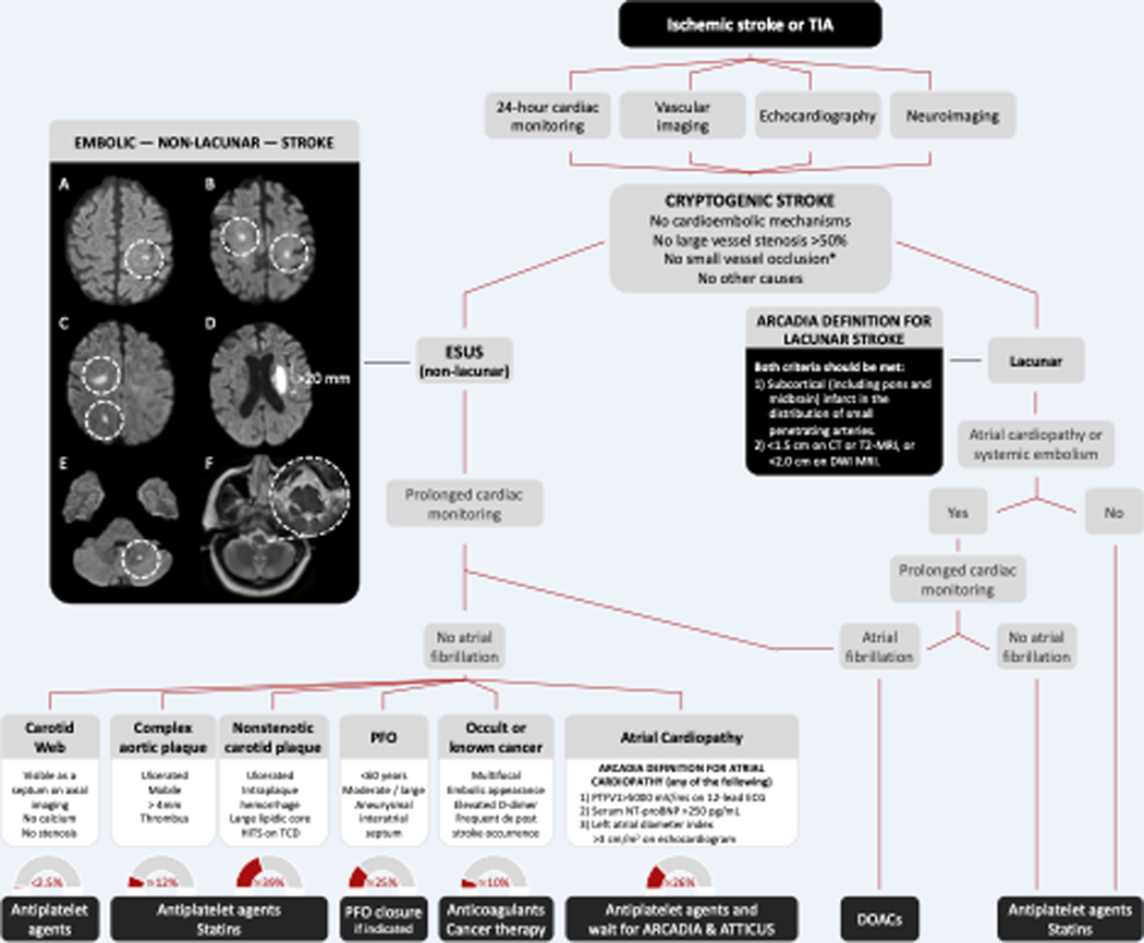
Figure 2. Proposed Algorithm for the Diagnosis and Treatment of Cryptogenic Stroke.
Panel A. Classical embolic stroke involving the cerebral cortex. Panel B. Bilateral small subcortical infarcts. Panel C. Small simultaneous subcortical infarcts involving the same vascular territory. Panel D. Single, large (>20 mm) deep infarct. Panel E. Embolic cerebellar infarct. Panel E. Lateral medullary infarct.
ARCADIA, AtRial Cardiopathy and Antithrombotic Drugs In prevention After cryptogenic stroke. ESUS, embolic stroke of undetermined source. CT, computed tomography. MRI, magnetic resonance imaging. DWI, diffusion-weighted imaging. PFO, patent foramen ovale. HITS, high-intensity transient signals. TCD, transcranial Doppler ultrasound.
No small vessel occlusion*, clinical TOAST criteria for small occlusion are a traditional lacunar syndrome and the absence of cortical deficits.

