
FIGURE 1.
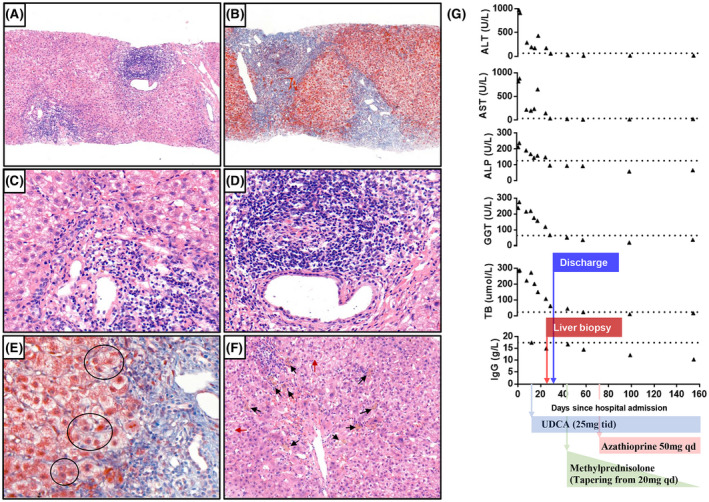
Histological findings and evolution of laboratory tests. At low‐magnification (×100), hematoxylin and eosin staining shows moderate to severe interface hepatitis with a dense lymphoid infiltrate (A), and Masson staining shows the formation of fibrous septa (B). At high magnification (×400) with hematoxylin and eosin stain, the interface necroinflammation consists primarily of lymphocytes with plasma cells (C); the dense periductal lymphocyte infiltrate (D) and the hepatic rosette formation (black circle) were well observed with Masson stain (E). Feathery degeneration of hepatocyte (red arrow) and hepatic cholestasis (black arrow) were also observed (F) (×200 hematoxylin and eosin stain). (G) Trends of liver function tests, total bilirubin, and total IgG levels over time. Dashed lines are the respective lower limit of the normal range of each test. ALP, alkaline phosphatase; ALT, alanine aminotransferase; AST, aspartate aminotransferase; GGT, gamma‐glutamyltransferase; TB, total bilirubin; UDCA, ursodeoxycholic acid