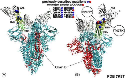
Figure 1.

Three‐dimensional structural representation of the SARS‐CoV‐2 Spike protein exhibiting the mutations present in Omicron variant receptor‐binding motif (RBM). Blue and red spheres represent the RBM's substitutions. nAb, vaccine‐induced neutralizing‐antibody (silver); RBM, receptor‐binding motif (yellow) and RBD, receptor‐binding domain (green). The image was created with the Visual Molecular Dynamics (VMD) v.1.9.3 (http://www.ks.uiuc.edu/Research/vmd/)
