Fig. 3.
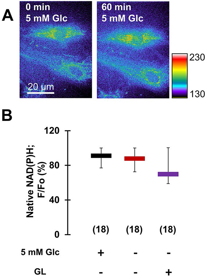
Aglycemia of astrocytes does not significantly affect glutamate oxidation in their mitochondria. A UV-induced autofluorescence imaging of native NAD(P)H in astrocytes (raw data) before (left), and after incubation of cells for 60 min (right) in 5 mM glucose (control, normoglycemia). The pseudo color scale is a linear representation of the fluorescence intensities ranging from 130 to 230 i.u.; scale bar, 20 μm. B Analysis of intracellular/mitochondrial NAD(P)H signal, reporting on oxidative metabolism, expressed as percentage of fluorescence retained (F/Fo) after the incubation period, shows no significant difference between NAD(P)H fluorescence in normoglycemic and aglycemic astrocytes or astrocytes lacking external glucose subjected to the GL protocol. All data points in B are shown as medians with IQRs. Number of astrocytes studied in each condition is given in parentheses in B. Other annotations as in Fig. 2

