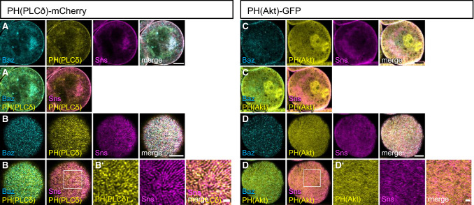
Fig. 1.
PI(4,5)P2 accumulates at slit diaphragms, whereas PI(3,4,5)P3 is rather found at the free plasma membrane. (A–D) Garland nephrocytes expressing either UAS::PH(PLCδ)-mCherry for labelling PI(4,5,)P2 or UAS::PH(Akt)-GFP to visualize PI(3,4,5)P3 were dissected from 3rd instar larvae, fixed and stained with the indicated antibodies. A and C are sections through the equatorial region of the nephrocyte; B and D are onviews onto the surface of these nephrocytes. Scale bars are 5 µm and 1 µm in insets

