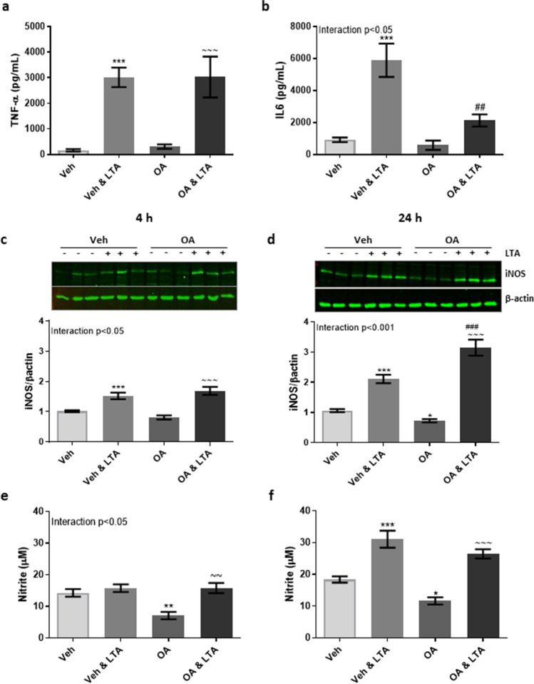
Fig. 3.
Oleic acid mitigates LTA-induced release of IL-6 and basal nitrite expression in microglia. BV2 cells were exposed to oleic acid (OA; 100 μM) or vehicle control (Veh) for 24 h, and stimulated with LTA (5 μg/mL) during the final 4 h, or for a further 24 h. Concentration of TNF-α (a) and IL-6 (b) was measured in the supernatant following 4 h LTA exposure (n = 6–45 replicates, from 3–12 independent experiments). Expression of iNOS and supernatant concentration of nitrite were examined both 4 h (c, e) and 24 h (d, f) following LTA stimulation (n = 9–45 replicates, from 3–12 independent experiments). Data is presented as mean ± SEM. *p < 0.05, **p < 0.01, ***p < 0.0001, compared to Veh; ~ ~ ~ p < 0.001, compared with OA; ###p < 0.001, compared with Veh + LTA. Interactions based on two-way ANOVA, followed by Bonferroni post-tests. Inserts illustrate representative immunoreactive bands for iNOS and β-actin (c, d; triplicate samples)

