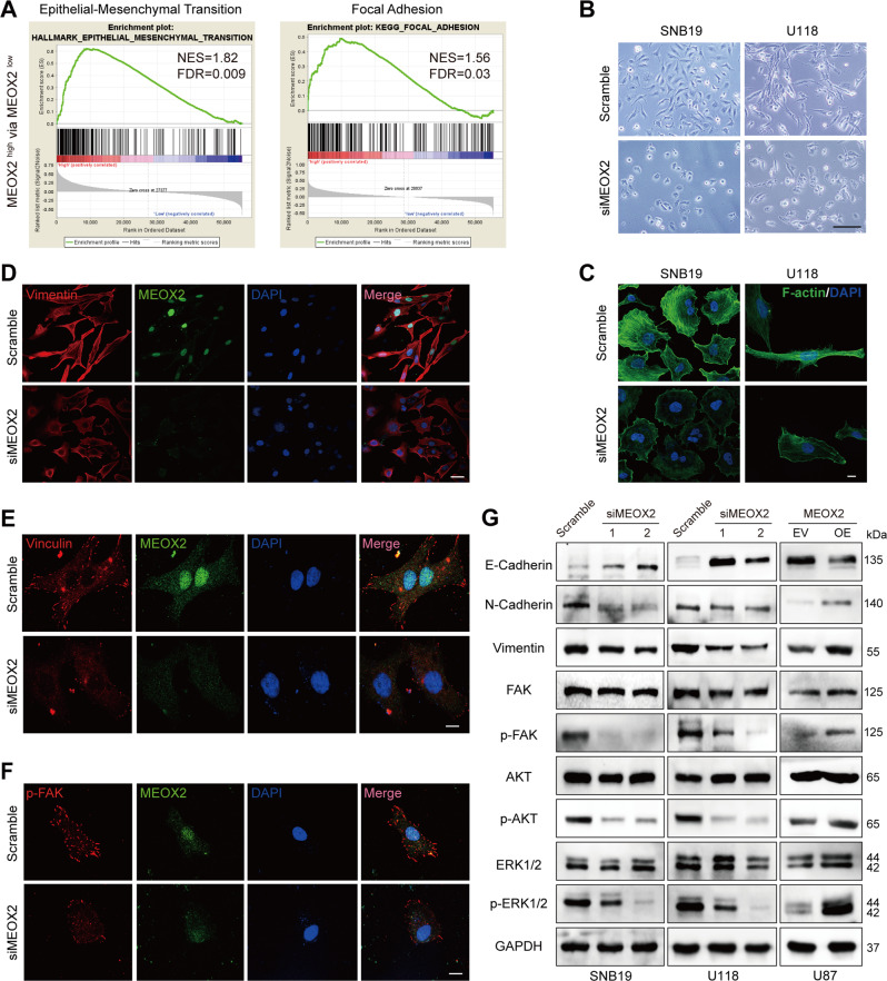
Fig. 4. MEOX2 mediates EMT process, formation of focal adhesion, and F-actin assembly in glioma cells.
A GSEA enrichment terms of EMT and focal adhesion in high MEOX2 expression vs low MEOX2 expression in TCGA gliomas. B The morphology of SNB19 and U118 cells treated with MEOX2 or Scramble siRNA was imaged by microscopy. Bar = 200 µm. C F-actin immunofluorescence staining of SNB19 and U118 cells with MEOX2 knockdown or control cells were observed by confocal microscopy. Bar = 200 µm. D–F Vimentin (D), Vinculin (E), and p-FAK (Y925) (F) immunofluorescence staining images of U118 cells with MEOX2 silencing or control cells were captured by confocal microscopy. Bar = 50, 100, 100 µm. G The protein expression of E-cadherin, N-cadherin, Vimentin, FAK, p-FAK (Y925), AKT, p-AKT (Ser473), ERK1/2, and p-ERK1/2 (Thr202/Tyr204) were analyzed by immunoblot in SNB19, U118 cells with or without MEOX2 inhibition and U87 cells with or without MEOX2 overexpression.

