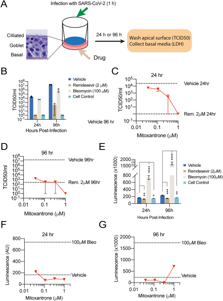
Figure 5.
Mitoxantrone inhibits SARS-CoV-2 infection in an EpiAirway 3D tissue model. (A) A schematic diagram of the experimental design. (B) Remdesivir (2 μM) but not Bleomycin (100 μM) inhibits SARS-CoV-2 infection. 24 or 96 h after drug treatment and viral infection (MOI 0.1), the cell surface was washed. The viral titer (TCID50) in the wash was determined. (C,D) Mitoxantrone inhibits SARS-CoV-2 infection in the EpiAirway 3D model. TCID50 was determined either 24 h (C) or 96 h (D) after the organoids were treated with the drug at the indicated concentrations and then air-infected with SARS-CoV-2 at MOI 0.1 for 1 h. The cells were washed from the apical side to remove the virus in the cell exterior and then incubated for 24 (C) or 96 h (D). Cells were rinsed from the apical side again and viral titers in the wash were determined. The dashed lines indicate the viral titer from cells infected without Mitoxantrone or in the presence of 2 mM Remdesivir (Rem.), as indicated. (E) Bleomycin (100 μM) but not Remdesivir (2 μM) induces cell death, releasing LDH as determined by a luciferase assay. *, p < 0.05, **, p < 0.01, ***, p < 0.001, ****, p < 0.0001 by unpaired student’s t-test. n = 2 tissues per test, each with 3 technical repeats. (F,G) Mitoxantrone inhibits SARS-CoV-2-induced cytotoxicity in the EpiAirway 3D model. As in (D,E) except that LDH in washes collected from Mitoxantrone-treated tissues was measured.

