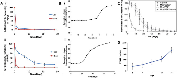
Figure 6.
(A) In vivo release profiles of (i) VEGF and (ii) BMP-2 were obtained after implantation of scaffold into the subcutaneous tissue mice for 28 days,303 where CSB and CSV represent nanocomposite fibrous scaffold (CS) loaded with BMP2 (B) and VEGF (V), respectively [Reproduced with permission from ref 303. Copyright 2018 Elsevier]. (B) Release kinetics of PLGA microspheres steady release of neurotrophic growth factors was detected for at least 60 days (i, BDNF) and 80 days (ii, GDNF) [Reproduced with the permission from ref 358. Copyright 2010 Springer Nature]. (C) Normalized in vivo release profile of BMP-2 from the four different implants in a rat subcutaneous implantation model, where Mps = PLGA microparticles loaded with BMP-2, and PPF = poly(propylene fumarate) [Reproduced with the permission from ref 359. Copyright 2008 Elsevier]. (D) Cumulative release of VEGF from the biohybrid scaffold with PLGA nanofibers, which displays a sustained release of VEGF over 28 days [Reproduced with the permission from ref 355. Copyright 2014 Elsevier].

