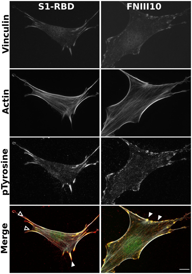
Figure 4. S1-RBD engagement initiates focal adhesion formation and actin organization.
FN−/− MEFs (2.5×10³ cells/cm2) were seeded on coverslips coated with 500 nM S1-RBD (left) or FNIII10 (right). Cells were incubated for 4 h prior to fixation and immunofluorescent staining for vinculin (green), actin (TRITC-phalloidin, white), or phospho-tyrosine (4G10, red). Arrowheads represent co-localization of vinculin and phosphotyrosine within focal adhesions (closed) and engagement with the actin cytoskeletion (open). Representative images shown from 1 of 4 independent experiments. Scale bar, 10 μm.

