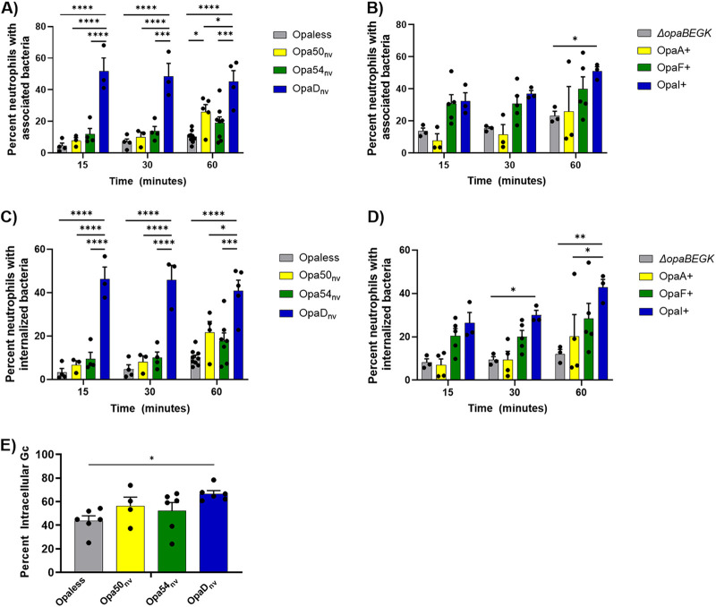
FIG 2.
Expression of different Opa proteins differentially affects binding and phagocytosis of N. gonorrhoeae by primary human neutrophils. The indicated strains of N. gonorrhoeae (A and C, constitutively expressed, nonvariable; B and D, phase variable) were labeled with Tag-IT Violet (TIV) and incubated with adherent, IL-8-treated primary human neutrophils. At the indicated times, cells were fixed and stained with DyLight 650 (DL650)-labeled anti-N. gonorrhoeae antibody without permeabilization to recognize extracellular bacteria. Neutrophils were analyzed via imaging flow cytometry. Panels A and B report the percentage of single, intact neutrophils with ≥1 cell-associated bacterium (TIV+). Panels C and D indicate the percentage of neutrophils with ≥1 phagocytosed bacterium (TIV+ DL650−). Results are the average of n ≥ 3 biological replicates. Data were analyzed by two-way ANOVA with Tukey’s multiple comparisons, with the following indications of significance: *, P < 0.05; **, P < 0.01; ***, P < 0.005; ****, P < 0.001. Only statistical comparisons within a time point were made. (E) The indicated strains of N. gonorrhoeae were labeled with CFSE and then incubated with adherent, IL-8-treated neutrophils. After 60 min, cells were fixed and stained with AlexaFluor 647 (AF647)-labeled anti-bacteria antibody without permeabilization. Images were captured by fluorescence microscopy. The percentage of intracellular N. gonorrhoeae was determined by dividing the number of CFSE+ AF647− (intracellular) N. gonorrhoeae by the number of CFSE+ AF647+ (total) N. gonorrhoeae. Statistical comparisons were made for n ≥ 4 biological replicates using one-way ANOVA with Tukey’s multiple comparisons, with P < 0.05 (*) considered significant.

