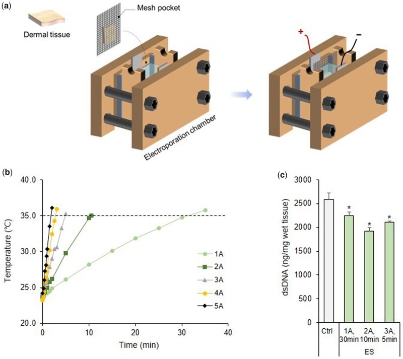
Figure 2.
(a) Schematic diagram of the electroporation system used for tissue decellularization. (b) Measurement of temperature change of 1 M NaCl while applying ES. (c) Quantification of the residual DNA content in skin tissues after ES under conditions where the temperature of the 1 M NaCl solution did not exceed 35°C. *P < 0.05 vs. Ctrl. dsDNA, double-stranded DNA

