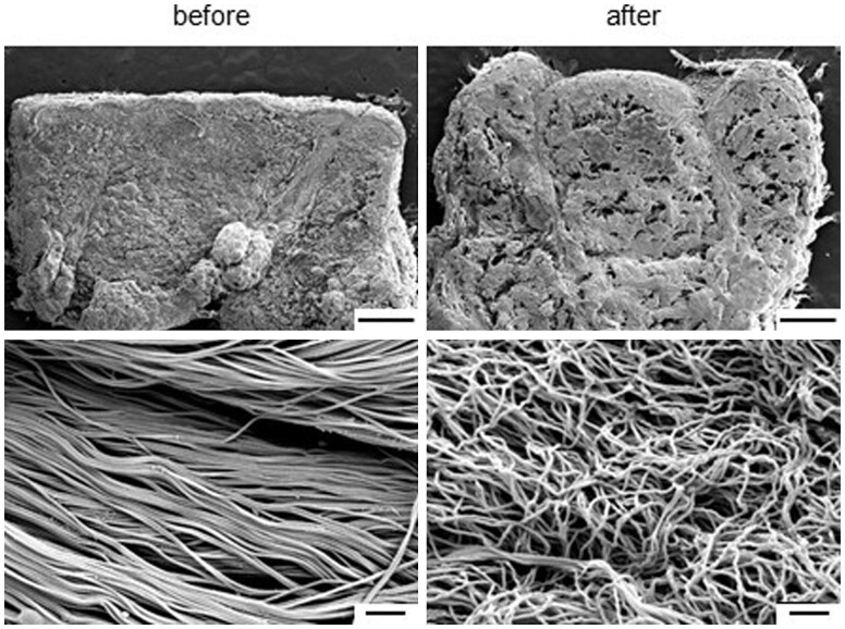
Figure 5.
Representative SEM images of the cross-section of skin tissues before and after immersion in the pretreatment solution. The upper image shows the presence or absence of the epidermal layer, and the lower image shows the dermis. Scale bar: 1 mm (upper) and 1 μm (bottom). SEM, scanning electron microscopy

