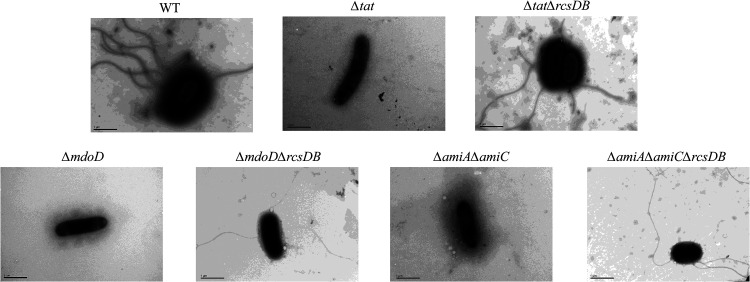
FIG 5.
Transmission electron microscopy analysis of flagella. Cells of each indicated strain were streaked on an LB agar plate at 37°C for 12 h. A single colony was picked and resuspended in 200 μL of ultrapure water, which was left to stand for 2 h. Then, 10 μL of the bacterial suspension was placed on the grid followed by fixing with 2% phosphotungstic acid staining solution and was imaged using a transmission electron microscope (H-7650; Hitachi, Japan). The scale bar is 1 μm.

