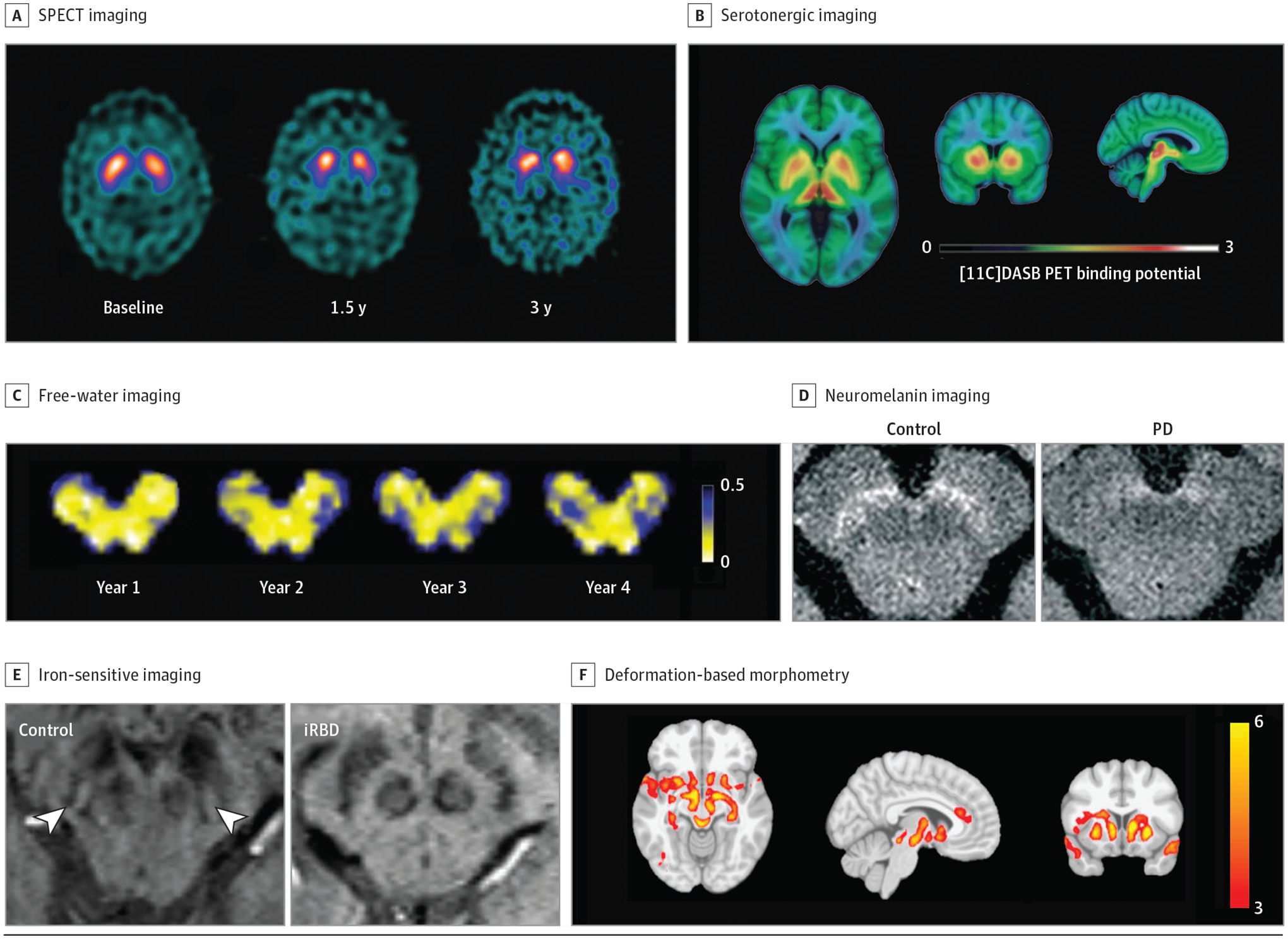
Figure 1.

Neuroimaging Biomarkers
Neuroimaging techniques are reviewed. A, Single-photon emission computed tomography (SPECT) imaging in controls and those with early-stage, moderate, and late-stage Parkinson disease (PD; reprinted with permission from Schapira and Olanow1). B, Positron emission tomography (PET) fluorodopa imaging in controls and those with early-stage and late-stage PD (reprinted with permission from Schapira and Olanow1). C, Free-water imaging longitudinally over 4 years in an individual with de novo PD (provided with courtesy of Roxanna Burciu, PhD [Department of Kinesiology and Applied Physiology, University of Delaware, Newark], and used with permission). D, Neuromelanin-sensitive imaging in a healthy control and patient with PD (disease duration of 4 years; reprinted with permission from Biondetti et al2). E, Absence of the nigrosome formation hypointensity using iron-sensitive imaging in a patient with idiopathic rapid eye movement sleep behavior disorder (reprinted with permission from De Marzi et al3). F, Distribution of atrophy in a patient with PD measured by deformation-based morphometry (reprinted with permission from Zeighami et al4).
