
Fig. 3.
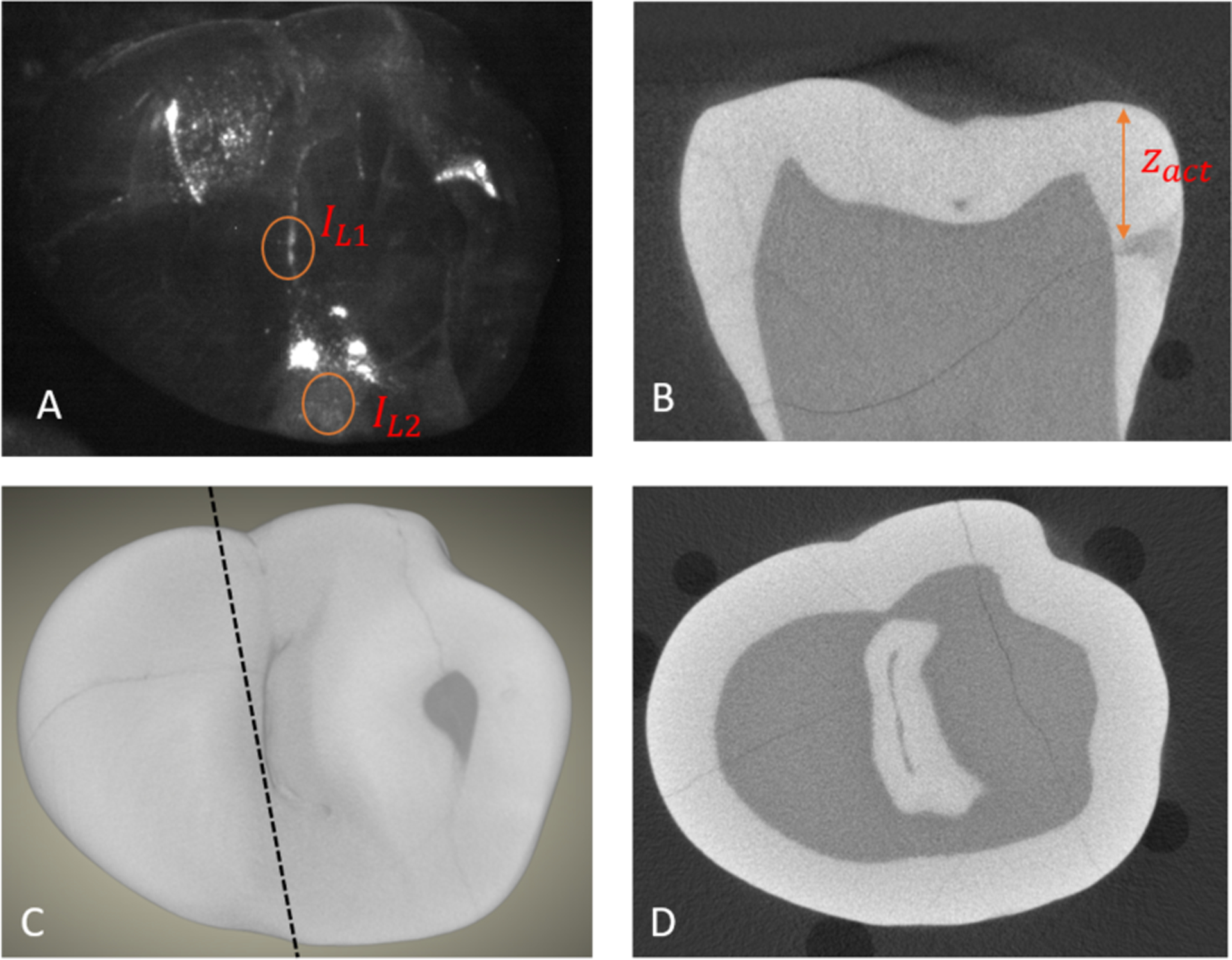
Estimating the depth from the occlusal surface of an interproximal lesion. (A) SWIR reflectance image of the tooth occlusal surface using a 2nd occlusal lesion for IL1 and the subsurface interproximal lesion intensity for IL2. (C) MicroCT surface rendering, the dashed line shows the position of the extracted slice shown in (B) that shows the distance between the tooth surface and the interproximal lesion (zactual). (D) MicroCT cross-section showing the occlusal surface lesion in the central fissure.