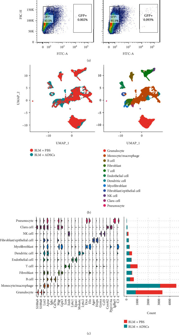
Figure 2.

Injected ADSCs greatly changes single-cell heterogeneity of BLM-treated lung tissue. (a) Flow cytometry diagrams for sorting of digested lung tissue based on GFP signal intensity. Left: BLM + PBS group, as non-GFP control for lung-originated cells only. Right: BLM + ADSCs group, showing a small population of GFP+ cells, which are considered recollected ADSCs. (b) UMAP plot of lung-originated cell scRNA-seq data, merging cells from the BLM + ADSCs group and BLM + PBS group. Left: dots colored by group. Right: dots colored by SingleR annotated cell type based on clusters identified by Seurat. (c) Stacked violin plot of cluster marker genes (three marker genes for each cluster) and cell count number for each cluster of lung-originated cells.
