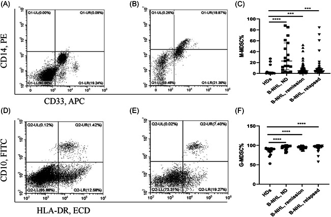
Figure 1.

(A) Representative flow cytometry plots of CD14 + CD33 + HLA‐DR−/low (M‐MDSCs) cells in healthy donors. (B) Representative flow cytometry plots of CD14 + CD33 + HLA‐DR−/low (M‐MDSCs) cells in B‐NHL patients. (C) M‐MDSCs in B‐NHL patients of ND, remission and relapsed compared to healthy controls. (D) Representative flow cytometry plots of CD10‐HLA‐DR−/low cells (G‐MDSCs) in healthy donors. (E) Representative flow cytometry plots of CD10‐HLA‐DR−/low cells (G‐MDSCs) in B‐NHL patients. (F) G‐MDSCs in B‐NHL patients of ND, remission and relapsed compared to healthy controls. *p < .05, **p < .01, ***p < .001, **** p < .0001, ns p ≥ .05. B‐NHL, B‐cell non‐Hodgkin lymphoma; G‐MDSC, granulocytic‐Myeloid‐derived suppressor cells; M‐MDSC, monocytic‐MDSC; ND, newly diagnosed
