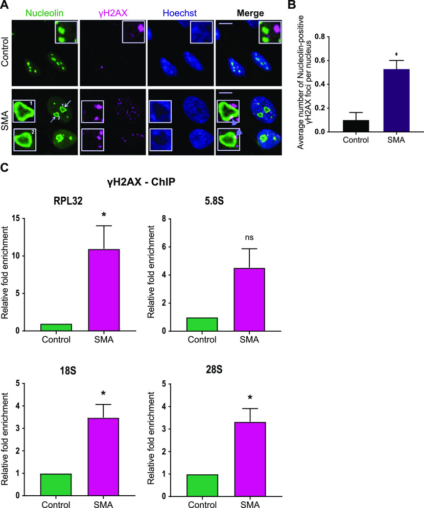
Figure 2. Survival motor neuron–deficient cells exhibit increased ribosomal DNA damage.
(A) Dual immunostaining with nucleolin and γH2AX performed on spinal muscular atrophy (SMA) type I and control fibroblasts. SMA type I fibroblasts form nucleolar caps (white arrows) that are shown to co-localise with γH2AX foci (violet arrowheads). Scale bars represent 5 μm. (B) Average number of nucleolin-positive γH2AX foci per nucleus. Data presented as mean ± SEM *P < 0.05, paired two-tailed t test (P = 0.0176). The data were collected from three biological independent replicates (N = 3). Nuclei counted = 50/replicate. (C) γH2AX-ChIP followed by qPCR analysis of ribosomal genes. Quantified RPL32, 5.8S, 18S and 28S gene qPCR data from γH2AX-ChIP experiment in SMA type I fibroblasts and healthy controls. IgG was used as a background control. Bar graphs of mean ± SEM (N = 3). *P < 0.05, ns, not significant (P > 0.05). Paired two-tailed t test; P = 0.0472 (RPL32), P = 0.0778 (5.8S), P = 0.0237 (18S), P = 0.0278 (28S).

