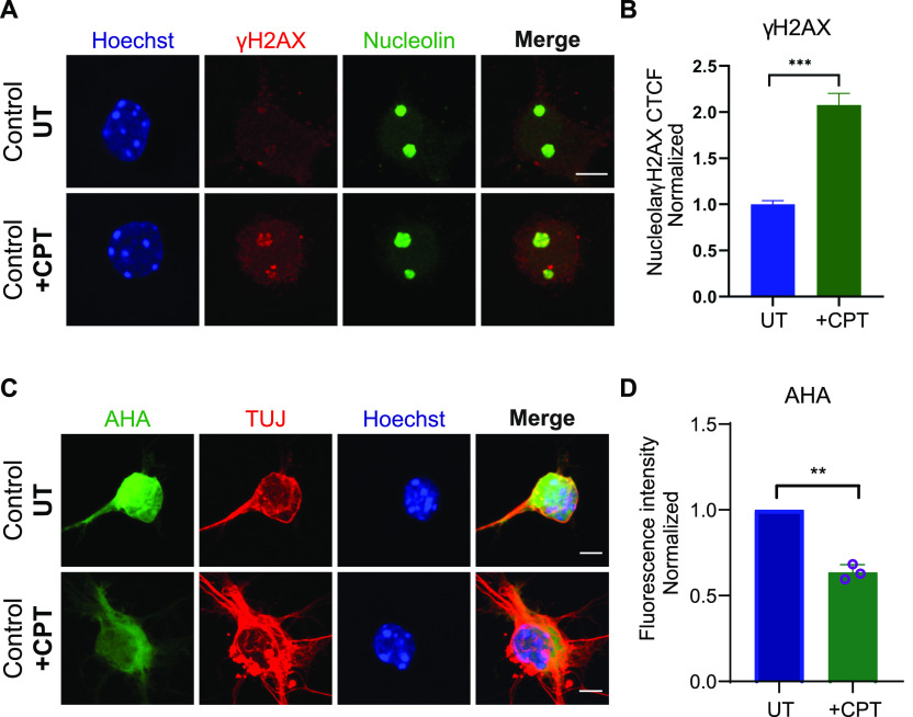
Figure 6. Extended exposure of control embryonic motor neurons to low levels of camptothecin (CPT) results in increased ribosomal DNA damage and subsequent translation impairment.
(A) Control wild-type embryonic motor neurons were treated with 50 nM CPT for 4 d. CPT-treated and untreated wild type embryonic motor neurons were double-stained with γH2AX (red) and nucleolin (green). Scale bars represent 5 μm. (B) Corrected fluorescence intensity of nucleolin-positive γH2AX signal was quantified. Briefly, for each replicate, we calculated the average value of all the UT points and used this as a normalizer. We then plotted all the normalised points from all three replicates in a single graph and performed Mann–Whitney non-parametric test. Bar graphs of mean ± SEM (N = 3). ∼150 cells analysed/replicate. ***P ≤ 0.001. (C) Protein synthesis was visualized by labelling newly synthesized proteins with AHA (green). Scale bars represent 10 μm. (D) AHA fluorescence intensity values in CPT treated cells normalised to untreated samples. Bar graphs of mean ± SEM (N = 3). **P < 0.01; paired two-tailed t test (P = 0.0049). Nuclei counted = 50/replicate.

