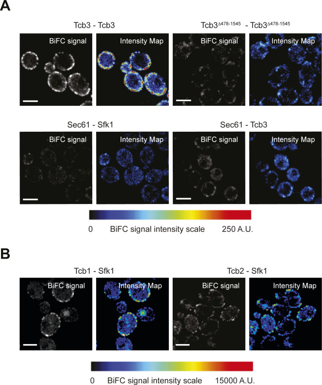
Figure S4. Tcb1 and Tcb2 associate with plasma membrane-localised Sfk1.
(A) Control protein–protein proximity assays; Protein–protein interactions between Tcb3 or Tcb3Δ478-1545 alone (top) or ER-localised Sec61 and either Tcb3 or Sfk1 (bottom) as detected by the split GFP BiFC assay. In each case, GFPN is fused to the protein listed on the left and GFPC is fused to the protein on listed on the right. (B) Protein–protein interactions between Sfk1 and Tcb1 (left) or Tcb2 (right).

