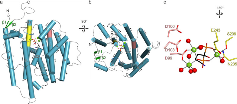
Fig. 3.
Overall architecture of one monomer of Copu9; a α-helices are drawn as blue cylinders and β-sheets as green arrows. The Asp-rich motif is colored in salmon and the NSE motif in yellow. The three Mg2+ ions are shown as green spheres and the AHD in stick representation; b View of panel a rotated by 90°, resulting in a view from the top into the active site; c detailed view of the Asp-rich and the NSE motif. The three Mg2+ ions are octahedrally coordinated by side chains of the catalytic motifs, water molecules and the phosphate functions of AHD

