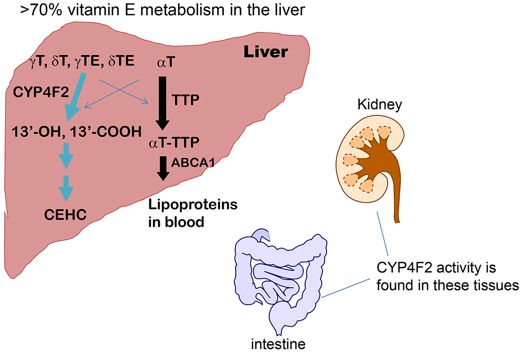
Fig. 3. Metabolism of vitamin E forms in the liver and possibly other tissues -.

In the liver, large portions of γT, δ;T, γTE and δTE are metabolized by CYP4F2-initiated ω-hydroxylation and oxidation to generate 13′-OHs and 13′-COOH, which is further metabolized to terminal metabolite CEHCs. In contrast, most αT and small amounts of other vitamin E forms are bound and transported by TTP (tocopherol transport protein) in hepatocytes and then incorporated into lipoproteins with the help of ABCA1 (ATP-binding cassette transporter A1). Vitamin E bound to lipoproteins are transported to other tissues via circulation. The crisscross arrows (light blue) indicate relatively minor events taking place for αT (catabolism) and other forms of vitamin E (binding to TTP) in the liver. It is estimated that over 70% metabolism of vitamin E takes place in the liver, while the kidney and intestine are found to have CYP4F2 activity and may therefore involve in vitamin E metabolism. (For interpretation of the references to colour in this figure legend, the reader is referred to the Web version of this article.)
