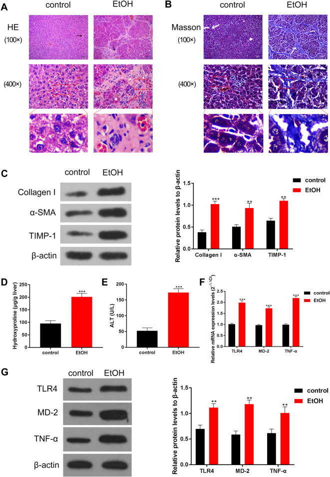
Fig. 1.
Alcohol treatment induced liver fibrosis and activated the TLR4/MD-2–TNF-α pathway in model rats.
In the alcohol-damaged liver of model rats, the molecules associated with liver fibrosis were upregulated, and the signaling pathway of liver fibrosis was activated. (A) Hematoxylin and eosin staining and (B) Masson staining were used to detect liver injury. The arrows indicate the portal area. Compared with that in the control group, the livers of rats in the EtOH group showed obvious pathological damage. (C) The levels of proteins related to liver fibrosis were detected by Western blotting, and data quantification was performed using ImageJ software; compared with that in the control group, the levels of proteins related to liver fibrosis in the EtOH group showed an obvious increase. (D–E) ELISA was used to determine the collagen-related markers (D) hydroxyproline and (E) ALT. The results showed that hydroxyproline and ALT levels significantly increased in the EtOH group. (F–G) The levels of TLR4, MD-2, and TNF-α were detected by (F) Western blotting and (G) RT-qPCR, and quantitation was performed using ImageJ software. The expression levels of TLR4, MD-2, and TNF-α were significantly increased in the EtOH group. The scale bar is 20 μm at high power and 80 μm at low power. **p<0.01, ***p<0.001, vs. the control group.

